Download
1 / 12
120 likes | 237 Views
This project focuses on the development of a cutting-edge tabletop X-ray microscope capable of providing high-resolution 3D imaging of living biological samples at the nanoscale. With a field of view of 10µm x 10µm and a resolution of 20 nm, this technology offers significant advantages over traditional imaging methods like fluorescence microscopy. The microscope will utilize advanced imaging techniques, including image stitching and X-ray tomography, to study dynamic biological processes in real-time, enhancing our understanding of cellular structures without the need for sample staining.

E N D
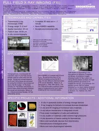
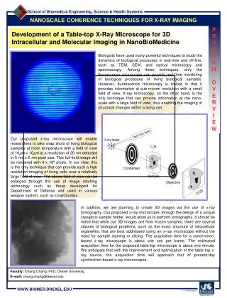
NANOSCALE COHERENCE TECHNIQUES FOR X-RAY IMAGING P R O G R A M O V E R V I E W Development of a Table-top X-Ray Microscope for 3D Intracellular and Molecular Imaging in NanoBioMedicine Biologists have used many powerful techniques to study the dynamics of biological processes in real-time and off-line, such as TEM, SEM, and optical microscopy and spectroscopy. Among these techniques, only the fluorescence microscope can provide real-time monitoring of biological processes of living biological samples. However, fluorescence microscopy is limited in that it provides information at sub-micron resolution with a small field of view. X-ray microscopy, on the other hand, is the only technique that can provide information at the nano-scale with a large field of view, thus enabling the imaging of structural changes within a living cell. Our proposed x-ray microscope will enable researchers to take snap shots of living biological samples at room temperature with a field of view of 10µm x 10µm at a resolution of 20 nm detected in 5 nm x 5 nm pixel size. This full-field image will be rendered with 4 x 106 pixels. In our view, this is the only technique that can provide such a high resolution imaging of living cells over a relatively large field of view. The above field of view can be enlarged through the use of image stitching technology such as those developed for Department of Defense and used in various weapon system, such as smart bombs. In addition, we are planning to create 3D images via the use of x-ray tomography. Our proposed x-ray microscope, through the design of a unique cryogenic sample holder, would allow us to perform tomography. It should be noted that while our 3D images are from frozen samples, there are several classes of biological problems, such as the exact structure of intracellular organelles, that are best addressed using an x-ray microscope without the need for sample staining or slicing. The acquisition time for a synchrotron-based x-ray microscope is about one sec per frame. The estimated acquisition time for the proposed table-top microscope is about one minute. We anticipate that with the improvement and optimization of the table-top x-ray source, the acquisition time will approach that of present-day synchrotron-based x-ray microscopes. • Faculty:Chang Chang, PhD, Drexel University. • E-mail: chang.chang@drexel.edu
Nanometer spatial resolution (~20nm) over 10 micrometer x 10micrometer field of view detected with 5nm x 5nm pixel size. NANOSCALE COHERENCE TECHNIQUES FOR X-RAY IMAGING P R O J E C T O V E R V I E W High Spatial and Spectral Resolution X-ray Microscope We expect to be able to achieve 10 frames of live biological samples before we exceed the threshold of radiation dosage that is tolerable for these biological samples. One strategy is to increase the number of frames after exceeding the allowable dosage to move to the previously unexposed neighboring region. • Faculty:Chang Chang, PhD, Drexel University. • E-mail: chang.chang@drexel.edu
X-ray microscope image of a human monocytic cell. This cell is imaged in suspension. NANOSCALE COHERENCE TECHNIQUES FOR X-RAY IMAGING P R O J E C T O V E R V I E W Human Monocytic Cell • Faculty:Chang Chang, PhD, Drexel University. • E-mail: chang.chang@drexel.edu
X-ray microscope image of a rat adrenal endothelial cell. The cells are cultured on a slab of thin (100nm) silicon nitride membrane, which also serves as a sample holder for the x-ray microsocpe. NANOSCALE COHERENCE TECHNIQUES FOR X-RAY IMAGING P R O J E C T O V E R V I E W Rat Adrenal Endothelial Cell • Faculty:Chang Chang, PhD, Drexel University. • E-mail: chang.chang@drexel.edu
NANOSCALE COHERENCE TECHNIQUES FOR X-RAY IMAGING P R O J E C T O V E R V I E W Estimated Exposure Time for Undulator-based X-ray Microscopes • Faculty:Chang Chang, PhD, Drexel University. • E-mail: chang.chang@drexel.edu
Sample Broadband Point source Photodiode NANOSCALE COHERENCE TECHNIQUES FOR X-RAY IMAGING P R O J E C T O V E R V I E W Optical Coherence Topography Our system, as designed, can hold biological samples of the order 5mm x 5mm with a sample thickness of about 10µm. Our sample positioning system would allow movements of 1mm in the x and y direction. This arrangement will provide us with 104 possible frames to look at. If we are studying biological processes that are taking place homogeneously across the sample, then we have 104 frames that can be exposed to a dosage below lethal level. Therefore, in theory, we have 104 x 10 snap shots to study the dynamic evolution of a biological process. This will enable us to take 1,400 snap shots within 24 hours. By reducing the frame acquisition time to 10 seconds, this number can be increased to about 8,000 frames. Considering the above performance specification of the x-ray microscope, there are large classes of biological problems involving structural changes at the nano scale that can only and uniquely be studied with this microscope, and we have identified several of these phenomena in this proposal (e.g., the Crabtree effect, mitochondrial structure change during rapoptosis, and nitric oxide transport mechanisms). • Faculty:Chang Chang, PhD, Drexel University. • E-mail: chang.chang@drexel.edu
Sample Extended Broadband Source CCD Grating 1 Grating 2 NANOSCALE COHERENCE TECHNIQUES FOR X-RAY IMAGING P R O J E C T O V E R V I E W Optical Sectioning • Faculty:Chang Chang, PhD, Drexel University. • E-mail: chang.chang@drexel.edu
Large depth-of-focus • Fluorescent friendly Condenser Microscope CCD NANOSCALE COHERENCE TECHNIQUES FOR X-RAY IMAGING P R O J E C T O V E R V I E W Microscopic Tomography • Faculty:Chang Chang, PhD, Drexel University. • E-mail: chang.chang@drexel.edu
- + Silicon nano fluidics channel Silicon nano array NANOSCALE COHERENCE TECHNIQUES FOR X-RAY IMAGING P R O J E C T O V E R V I E W Automated High Throughput Platform • Faculty:Chang Chang, PhD, Drexel University. • E-mail: chang.chang@drexel.edu
NANOSCALE COHERENCE TECHNIQUES FOR X-RAY IMAGING P R O J E C T O V E R V I E W Holographic Imaging • Faculty:Chang Chang, PhD, Drexel University. • E-mail: chang.chang@drexel.edu
Broadband Point Source Sample Photodiode NANOSCALE COHERENCE TECHNIQUES FOR X-RAY IMAGING P R O J E C T O V E R V I E W Holographic Imaging Schematic • Faculty:Chang Chang, PhD, Drexel University. • E-mail: chang.chang@drexel.edu
NANOSCALE COHERENCE TECHNIQUES FOR X-RAY IMAGING P R O J E C T O V E R V I E W Simulation of the XOR Pattern Conclusion The ultimate goal of this proposal is to make the experience and know-how generated in the proposed activity easily duplicated for any regional user's facility for replication of their own table-top system, thereby making the power of x-ray microscopy available to a wider user base. • Faculty:Chang Chang, PhD, Drexel University. • E-mail: chang.chang@drexel.edu