Download
1 / 17
180 likes | 838 Views
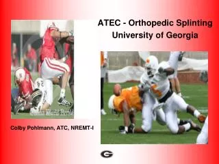
Colby Pohlmann, ATC, NREMT-I. ATEC - Orthopedic Splinting University of Georgia. Traction Splint Application. NOTE : Is to be used only for a painful, swollen, deformed mid thigh injury with NO lower leg injury.

E N D
Colby Pohlmann, ATC, NREMT-I ATEC - Orthopedic Splinting University of Georgia
Traction Splint Application NOTE: Is to be used only for a painful, swollen, deformed mid thigh injury with NO lower leg injury. This information is designed to be used as a guide for an “Ischial” type traction splint. There are several different types of commercially made traction splints available. This information may differ for the device that you use.
Why a Traction Splint? The theory behind the traction splint is that it reduces potential blood loss by separating and aligning the fracture segments through traction. This serves to keep the thigh at its normal length and relatively normal circumference - thus decreasing the potential space for blood loss.
Contraindications for the use of a Traction Splint 1 - Partial amputation or avulsion with bone separation, or the distal limb is connected only by marginal tissue. 2 - Injury is close to the knee 3 - Injury to the knee 4 - Injury to the hip 5 - Injury to the pelvis 6 - Lower leg or ankle injury
Application of the Traction Splint 1 - Take appropriate body substance isolation precautions. 2 - Apply manual stabilization Apply manual stabilization to the leg above and below the injury site. This is designed to stabilize the bone ends and reduce further injury. 3 - Explain the procedure to the patient The athlete may be very anxious about this procedure. You need to properly communicate to the athlete what you will be doing. 4 - Remove clothing from the area Remove the clothing to expose the entire leg, then remove the shoe and sock from the effected extremity.
Traction Splint Cont. 5 - Assess pulse, motor, and sensory function distal to the injury and compare to the opposite (non-injured) extremity. 6 - Apply the ankle hitch After the ankle hitch is in place, elevate the leg while supporting the ankle. 7 - Measure the traction splint Adjust the traction splint to the proper length. The non-injured leg should be used to measure the length of the traction splint. The traction splint should be adjusted to 12 inches longer than the non-injured leg.
Traction Splint cont. 7 - Apply the traction splint Slide the traction splint under the patient’s injured leg, the ischial ring of the traction splint must be against the bony prominence of the ischial tuberosity. If equipped with a kickstand at the end of the traction splint, extend it once the traction splint is in place. Pad the groin and gently, but securely apply the ischial strap. You should be able to fit two fingers between the ischial strap and the patient’s thigh to prevent over tightening.
Traction Splint cont. 8 - Apply mechanical traction Attach the mechanical traction device to the ankle hitch. Avoid using too much traction, which may overstretch the leg, but use enough traction to maintain limb alignment. Many patients will have reduced pain and muscle spasms once adequate mechanical traction is applied. 9 - Secure the leg to the traction splint Fasten the series of support straps. One strap should be just above the ankle hitch, one strap just below the knee, one strap just above the knee, and one strap at the top of the thigh just below the ischial strap. Do not fasten a strap directly over the injury site. Excess straps should be secured underneath the splint to provide additional support.Recheck the ischial strap to assure that it has not loosened. 10 - Reassess distal pulses, motor, and sensory function distal to the injury site and compare to the opposite non injured extremity. 11 - Prepare the patient for transport The patient should now be secured to a long backboard to provide further immobilization of the hip. The traction splint should also be secured to the long backboard to prevent excessive movement.
Using the Sam Splint • Check PMSC and control major bleeding. • Shape the splint to the limb. You’ll want to immobilize the joint above and the joint below the injury. With the example of a forearm injury, the splint extends below the wrist (immobilizing it) and above the elbow (immobilizing it). Make no attempt to straighten a suspected fracture while using this splint. Splint it exactly as it’s found. • Bend the splint into a U-shape. This cradles the arm, giving greater protection and making the splint more comfortable. It also give the splint greater structural strength.
Sam Splint cont. • Wrap the splint and the limb with a roller bandage so that the splint and the limb are firmly bonded together. Don't make the wrapping so tight that blood flow through the limb is obstructed. Commonly-used wrapping materials include Coban, Ace Bandages, Roll gauze, and Adhesive tape. • For upper extremity injuries, place a sling on the patient to keep the arm elevated and immobile. A chest strap across the arm in a sling will keep the arm tight against the chest.
When securing the splint to the limb, remember that you need to keep an open area for monitoring pulse, motor, sensation and circulation. For open fractures or other open wounds, the application of the splint is the same. However, you may need to apply sterile bandages or dressings to the open wounds before placing the splint in place. For lower extremity applications, you may need to use two splints instead of one. Two splints can be overlapped at one end and taped in place with adhesive tape. To increase structural strength, after curving the splint in a "U" shape, bend the edges down slightly. FYI
Rapid Form Immobilizer • Assess the pulse, motor, sensation and circulation of the injured area. • For splinting to be effective, the joints above and below the fracture must be immobilized. • If possible, remove any clothing that may impede the splint's ability to work properly. • If there are open wounds or exposed bone, bandage appropriately. • The injured area must be manually stabilized, which prevents movement. This can be done by simply holding the affected area, preventing movement above and below it. For example, for a radius/ulna fracture, the arm should be held at the wrist and elbow.
Rapid Form Immobilizer • When using vacuum splints, place the injured extremity inside the splint. • Use the pump to draw air out of the splint, which compresses it, making it rigid. It also conforms to the patient and reduces pressure on the area. • When using vacuum splints, make sure to keep the patient's fingers and/or toes exposed to assess motor function and capillary refill. • The splint should be checked periodically during transport to ensure there are no leaks. Leaks in the splint diminish its rigidity and effectiveness.
Kendrick Extrication Device The Kendrick Extrication Device (KED) is designed to immobilize a patient found in a sitting position. It is most commonly used in automobile accidents where the patient is stable. If the patient is unstable, you will need to perform a Rapid Extrication.
Procedural Protocols • Rescuer One should be positioned behind the patient to stabilize the head and neck. • 2. Rescuer Two checks neurological and vascular response of all • extremities. • 3. Rescuer Two measures and applies the cervical collar. • 4. The KED is slide into position behind the patient.
Procedural Protocols cont. 5. The KED is wrapped around the patient, and the middle strap is secured. (The KED should be snug beneath the patient’s armpits) 6. The bottom strap is secured next. 7. The top strap of the KED is secured. 8. Each leg strap is wrapped around the leg and secured. 9. The patient’s head is secured into the KED.
Procedural Protocols cont. 10. All of the straps are tightened down. 11. The patient’s wrist and legs are secured. 12. A long spine board is placed under the patient’s buttocks. 13. Remove patient from the vehicle and transferred to the spine board. 14. Disconnect the leg straps, allowing the patient’s legs to lay flat on the long spine board. 15. Refer to the securing a patient to the long spine board. *** Reminder *** - Neurological and vascular checks should be performed on the patient prior to and after extrication. -If the patient’s becomes unstable at any time, refer to a Rapid Extrication Protocol.