Download
1 / 48
480 likes | 698 Views
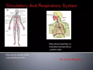
The Respiratory System and The Circulatory System. Prepared By: Chris Reyes, Robert Rock and Keturah Reed. The Circulatory System. The Circulatory System utilizes the heart to circulate blood within the body using blood vessels. Heart Healthy.

E N D
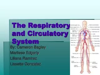
The Respiratory System and The Circulatory System Prepared By: Chris Reyes, Robert Rock and Keturah Reed
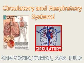
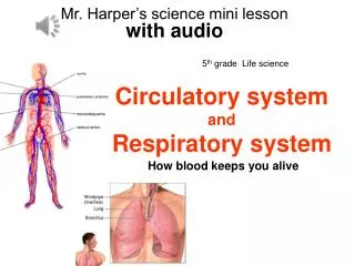
The Circulatory System • The Circulatory System utilizes the heart to circulate blood within the body using blood vessels.
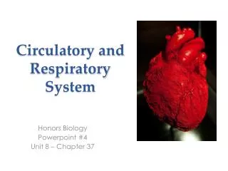
Heart Healthy • The heart is made up of four chambers - two atria and two ventricles. The atria receive blood returning to the heart while the ventricles pump blood from the heart. • The heart is the muscle that works to pump blood oxygenated throughout the body through arteries and receives deoxygenated blood through veins. The heart is located beneath the sternum and is about the size of a fist. It consists mostly of cardiac muscle. The vena cava is the vein that directly brings blood back to the heart. The two atria have relatively thin walls and serve as collection chambers for returning blood. Contraction of the atria completes filling of the ventricles. • (Holmes, 2004) (Cambell & Reece, 2005) (Scogna 2004)
The ventricles have thicker walls and contract more strongly. The left ventricle contract very strongly because it is responsible for pumping blood through the aorta to al parts of the body. The heart contracts on a rhythmic cycle. Contraction pumps blood and when the hearts relaxes, chambers fill with the blood. the cardiac cycle refers to the complete sequence of pumping and filling. The contraction of blood is the heart beat. The relaxation phase is the diastole and the systole is the contraction phase. The hydrostatic pressure that blood exerts against the walls of a vessel and that propels the blood is blood pressure. (Holmes, 2004) (Cambell & Reece, 2005) (Scogna 2004)
Cardiac output is the volume of blood the left ventricle pumps out per minute. four valves in the heart prevent backflow of blood and blood from moving in the wrong direction. Atrioventricular valves are between each atrium and ventricle. Semilunar valves are located at the two exits. (Holmes, 2004) (Cambell & Reece, 2005) (Scogna 2004)
The pulse is the rhythmic stretching of arteries caused by the pressure of blood driven by powerful contractions of the ventricles which can measure heart rate. The sinoatrial node, is the pacemaker of the heart and sets the rate and timing at which all cardiac muscle cells contract. The SA node creates electrical impulses that cause both atria to contract in unison. Impulses also pass through to atioventrical node which ensures that the atria empty completely before ventricles. These impulses can be conducted through an electrocardiogram. • (Holmes, 2004) (Cambell & Reece, 2005) (Scogna 2004)
Circulation • The body's circulatory system has three distinct parts: Pulmonary circulation, coronary circulation, and systemic circulation. • Coronary circulation refers to the movement of blood through the tissues of the heart. • Pulmonary Circulation refers to the movement of blood to and from the lungs • Systemic circulation supplies nourishment to all of the cells located in the body (with the exception of the lungs and the heart because they have their circulation systems) through the aorta • (Holmes, 2004) (Cambell & Reece, 2005) (Scogna 2004)
Pulmonary circulation begins as the right ventricle pumps blood to the lungs via the pulmonary arteries. As the blood flows from the capillary beds, in the left and right lungs, it loads oxygen and unloads carbon dioxide attained from cellular work. the oxygen rich blood flows into the left ventricle as the ventricle opens and the atrium contracts. The left ventricle pumps oxygen rich blood through the systemic circuit. Systemic circulation supplies nourishment to all of the cells located in the body (with the exception of the lungs and the heart because they have their circulation systems) through the aorta. The blood vessels (arteries, veins, and capillaries) are responsible for the delivery of oxygen and nutrients to the tissue. The forceful contraction of the heart's left ventricle forces the blood into the aorta which then branches into many smaller arteries which run throughout the body. The inside layer of an artery is very smooth, allowing blood to flow quickly. The outside layer of an artery is very strong, allowing the blood to flow very forcefully. Veins are thinner walled and convey blood back to the heart and flow back to the heart as a result of muscle action. (Holmes, 2004) (Cambell & Reece, 2005) (Scogna 2004)
Many capillaries are located in every tissue to ensure every part of the body is supplied with blood. The distribution of blood in the capillaries depends on smooth muscles controlled by the nervous system and hormones. It is through capillaries that the critical exchange of substances between blood and interstitial fluid occur. The waste products are collected and waste rich blood flows into the veins which bring bloods back to the heart. at the heart, through pulmonary circulation, gas exchange occurs in the lungs. During systemic circulation, blood passes through the kidney where much of the waste is filtered out. Blood also collects in the portal vein of the small intestine, where it passes through the liver, which filters sugars from the blood and stores them for later. (Holmes, 2004) (Cambell & Reece, 2005) (Scogna 2004)
Blood is made up of different cells suspended in plasma. Blood plasma is 90% water but also consists of inorganic salts, plasma proteins, and substances in transit. These in transit substances include nutrients, metabolic wastes, respiratory gases, and hormones. • The cellular elements of blood make up 45% of composition. Erthrocytes are red blood cells. They transport oxygen and help transport carbon dioxide. • Leukocytes are white blood cells that are responsible for defense and immunity. There are four types: Basophils, Neutrophils, Lymphocytes, Monocytes, and Eosinophils. Platelets are also cellular elements that aid in blood clotting. • (Holmes, 2004) (Cambell & Reece, 2005) (Scogna 2004)
Take Care… • Many complications can arise from improper care of the cardiovascular system. Cardiovascular diseases are diseases of the heart and blood vessels. • Lifestyle plays a major role to the development of cardiovascular diseases. Cholestrol is a major contributor. It travels in blood plasma as LDLs and HDLs. LDLs are low-density lipoproteins are bad and are associated with deposition of cholesterol plaque in the inner walls of arteries. • HDLs are high-density lipoproteins that reduce cholesterol deposits on arteries. Atherosclerosis (constant plaque buildup in arteries), hypertension (high blood pressure), heart attack (death of cardiac muscle tissue resulting from prolonged blockage of one or more of the coronary arteries), and stroke ( death of nervous tissue in brain from blockage or rupture of arteries) are problems that can result from complications with the cardiovascular system. • (Holmes, 2004) (Cambell & Reece, 2005) (Scogna 2004)
Breathing It In – The Respiratory System • The Respiratory System is made up of the nasal cavity and pharynx, larynx, lungs, trachea, and bronchi. Its main function is to supply the body with oxygen and take away carbon dioxide. Breathing is so automatic to life that we don’t even have to think about it. Each day we take 20,000 breaths. • (Holmes, 2004) (Cambell & Reece, 2005) (Scogna 2004)
With every breath, we take in oxygen-rich air through our nose and mouth and our lungs fill up and empty out. Even if the air we breathe is dirty or polluted, our respiratory system can defend itself through foreign substances that enter through the nose or mouth. Pollutants are breathed out again, coughed up, swallowed, passed out through the intestines or destroyed by digestive juices, or eaten by macrophages, a type of blood cell that patrols the body looking for germs to destroy.
The Journey of Air • In the nostrils air is taken in warmed and humidified. Cilia are tiny hairs that protect the nasal passageways and other parts of the respiratory tract. They filter out dust and other particles that enter the nose through breathed air. Air can also be taken in through the mouth. Air travels down the pharynx to the larynx. When eating, the larynx is covered by the epiglottis – a small flap of tissue that keeps food and liquid going into the larynx into the lungs. The larynx is adapted as a voice box. Sounds are created when voluntary muscles in the voice box or tensed. • (Holmes, 2004) (Cambell & Reece, 2005) (Scogna 2004)
Next air travels down the trachea (windpipe). The walls of the trachea are strengthened by stiff rings of cartilage which keep it open. The trachea is also lined with cilia, which keep fluids and foreign particles out of the airway so that they stay out of the lungs. Finally, the trachea divides into two right and left tubes known as bronchi. The bronchi connect to the lungs. In the lungs, the bronchi branch into smaller bronchi and even smaller bronchioles which end in tiny air sacs called alveoli. It is at alveoli that the exchange of oxygen and carbon dioxide takes place. Oxygen diffuses into a web of surrounding capillaries, while carbon dioxide diffuses in the opposite direction from the capillaries into the air space. (Holmes, 2004) (Cambell & Reece, 2005) (Scogna 2004)
Each lung has about 300 -400 million alveoli. The lungs contain elastic tissues that allow them to inflate and deflate without losing their shape and are encased by a thin lining known as the pleura. • The thorax (chest cavity) houses the bronchial tree, heart, lungs and other structures. The ribs and attached muscles form the top of the thorax and the bottom is formed by a large muscle – the diaphragm. • (Holmes, 2004) (Cambell & Reece, 2005) (Scogna 2004)
We use negative pressure breathing for respiration. Our respiratory system works as a pump, pulling air into the lungs. Diaphragm contracts and moves down when air is inhaled. The rib cage espands as rib muscles contract. When air exhaled, the diaphragm relaxes and moves up. The rib cage gets smaller as the rib muscles relax. Since the lungs do not completely empty and refill with each breath cycle, newly inhaled air mixes with oxygen-depleted residual air, and the maximum oxygen concentration in the alveoli is much less than that in the atmosphere.
Breathing in humans is controlled by the breathing control centers which are both located in the brain – the medulla oblongata, and the pons. The breathing control center in the medulla sets the basic rhythm for breathing and the pons moderates it. (Holmes, 2004) (Cambell & Reece, 2005) (Scogna 2004) The medulla has nerves that sends impulses to the diaphragm and the rib muscles causing them to contract and inhale. The medulla’s control center also aids in controlling the level of carbon dioxide in blood. The sensors in the medulla detect changes in pH of blood and cerebrospinal fluid soaking the surface of the brain. There are other sensors in the carotid arteries in the neck that can detect changes in blood pH and sends messages to the medulla. The medulla is able to alter breathing accordingly.
Respiratory pigments are proteins that transport oxygen and carbon dioxide and help buffer blood. They increase the amount of oxygen blood can carry. Hemoglobin is the respiratory pigment humans use. Besides assisting in oxygen and carbon dioxide transport, it also helps ensure no harmful pH changes occur in blood.
Reference Page • Scogna, Kathleen.(2004). Respiration. In K. Lee Lerner and Brenda Wilmoth Lerner (Ed.), Gale Encyclopedia of Science.3rd ed. Detroit: Gale, 2004. Retrieved April 30, 2008 from Science Resource Center. Thomson Gale. fi.edu/learn/heart/systems/respiration.html > • Campbell, N.A. & Reece, J.B.(2005). Carbon and the Molecular Diversity of Life. In B. Wilbur (Ed.), AP Edition Biology (7th ed.)(pp. 867 - 896). San Francisco: Benjamin Cummings • Holmes, Leonard D. “Circulatory System." Gale Encyclopedia of Science. Ed. K. Lee Lerner and Brenda Wilmoth Lerner. 3rd ed. Detroit: Gale, 2004. Science Resource Center. Thomson Gale. 14 November 2007 <library.thinkquest.org/5777/cir1.htm >
Activity 1 Lymphocyte Detective
You are T. Lymphoson, a detective in the white blood cell police department responsible for taking care of Peter Jameson’s circulatory system. • Peter is a middle-aged man recovering from prostate cancer. He has just been released from the hospital after undergoing surgery to remove early developing tumors in his prostate. • The police force has been extremely lethargic due to the extensive bed rest after the surgery but is on high alert for any signs of disease that might be caused by a metastatic tumor. • You, Detective Lymphoson, are responsible for patrolling the circulatory system for any suspects who might cause harm to Peter.
Urgent Situation!! • The blood pressure in Peter’s right leg has risen, possibly due to a blockage in one of the blood vessels there. He is in extreme pain and there is swelling in the muscles of his thigh. • Detective Lymphoson suspects it is plaque buildup in the arteries due to Peter’s unhealthy high cholesterol diet. He uses his map of the major blood vessels of the leg to navigate through the leg and round up suspects.
On The Trail • Detective Lymphoson must question red blood cells, platelets, and other white blood cells in the leg to find out if they have seen something. • The high blood pressure can be the cause of some sickness resulting from Peter’s weakened immune system after the cancer surgery. • Use these websites to find possible causes of high blood pressure and swelling in the body: http://www.americanheart.org/presenter.jhtml?identifier=2152 http://www.mayoclinic.com/health/high-blood-pressure/HQ01345
Culprit Found • The source of the pain and cause of the pressure has been found. It is a thrombus in the great saphenous vein. • Detective Lymphoson calls for backup but it is no use, the circulatory police have nothing that can dislodge the thrombosis. As the pain and swelling worsen Peter goes to the hospital.
At the Hospital • Peter is diagnosed with Thrombophlebitis due to his extensive bed rest after the operation. He is given anticoagulant medications which should break up the clot and clear the vein. • Use the web resources to research thrombophlebitis and find possible anticoagulant medications that would be the best fit for Peter’s condition: http://www.mayoclinic.com/health/thrombophlebitis/DS00223 http://www.healthatoz.com/healthatoz/Atoz/common/standard/transform.jsp?requestURI=/healthatoz/Atoz/ency/anticoagulant_and_antiplatelet_drugs.jsp
A New Ally • The anticoagulant medicine, Warfarin, is administered. It spreads throughout the body and thins Peter’s blood. • The thrombus is dislodged but the problem is not completely over. Peter’s blood clot could become lodged in other blood vessels. • It is your job to create a diet and medication plan that will prevent blood clots and lower Peter’s blood pressure. • Use these websites and others of your choice to create a plan of action: http://yourtotalhealth.ivillage.com/blood-clots.html http://www.cdc.gov/bloodpressure/prevention.htm
Level 1- Heart Healthy • What is the main function of the heart? • Name the four chambers of the heart and their function. • What seperates these chambers and what prevents the backflow of blood?
Level 1 - Answers • The heart’s main function is to receive deoxygenated blood, direct it to the lungs and pump oxygenated blood thrughout the entire body. • The four chambers of the heart are the right atrium, right ventricle, left atrium and left ventricle. • The heart is the size of a clenched fist. • GOOD JOB!! If you answered accurately, advance to level 2. If not, go back and read the introduction, because you have been eliminated. I guess a blood cell can beat you anyday…
Name the three types of circulation and their functions. • What is cholestrol? Name the types and how it travels.
The body's circulatory system has three distinct parts: Pulmonary circulation, coronary circulation, and systemic circulation.Coronary circulation refers to the movement of blood through the tissues of the heart. Pulmonary Circulation refers to the movement of blood to and from the lungs. Systemic circulation supplies nourishment to all of the cells located in the body (with the exception of the lungs and the heart because they have their circulation systems) through the aorta. • Cholestrol is travels in blood plasma as LDLs and HDLs. LDLs are low-density lipoproteins are bad and are associated with deposition of cholesterol plaque in the inner walls of arteries. HDLs are high-density lipoproteins that reduce cholesterol deposits on arteries. • Doing GREAT!! Keep up the good work!
Level 3 • What does blood carry that is essential to cellular work and how does it transport this? • What does blood consist of? • Name the types of white blood cells and their function.
Blood carries oxygen through the respiratory pigment, hemoglobin. • Blood is made up of different cells suspended in plasma. Blood plasma is 90% water but also consists of inorganic salts, plasma proteins, and substances in transit. These in transit substances include nutrients, metabolic wastes, respiratory gases, and hormones. • The cellular elements of blood make up 45% of composition. Erthrocytes are red blood cells. They transport oxygen and help transport carbon dioxide. • Leukocytes are white blood cells that are responsible for defense and immunity. There are four types: Basophils, Neutrophils, Lymphocytes, Monocytes, and Eosinophils. Platelets are also cellular elements that aid in blood clotting.
Breathing Made Easy • What makes up the respiratory system • Name the function of the respiratory system.
The Respiratory System is made up of the nasal cavity and pharynx, larynx, lungs, trachea, and bronchi. Its main function is to supply the body with oxygen and take away carbon dioxide.
What happens to inhaled pollutants? • Do we have a positive pressure breathing system. • How does our system function to inhale air?
Pollutants are breathed out again, coughed up, swallowed, passed out through the intestines or destroyed by digestive juices, or eaten by macrophages, a type of blood cell that patrols the body looking for germs to destroy. • We use negative pressure breathing for respiration. Our respiratory system works as a pump, pulling air into the lungs. Diaphragm contracts and moves down when air is inhaled. The rib cage espands as rib muscles contract. When air exhaled, the diaphragm relaxes and moves up. The rib cage gets smaller as the rib muscles relax.
It’s Getting Harder & Harder to Breathe Activity 3
Doctor Parrish M.D. • You are Dr. Leonardo Parrish the leading professional on respiratory disorders. • The patients name is Anastasia Lamari, she has been rushed to the hospital on what seems to be a severe case of asthma. • Along with your resident doctors Michaelangelo, Donatello, and Raphael you must discover what plagues Ms. Lamari.
Background of Ms. Lamari • Ms. Lamari is a 27 year old women that leads a very active life. She has had asthma since she was born, but it has never really taken a toll on her. The only time of seriously known problems is when her asthma occurs during the allergy season. Before the hospital she was running in the park with her friend when suddenly she felt the shortness of breath attack her and suddenly she collapsed. • Your job is to discover if it was just allergies with asthma which caused her fall or if it was something more...
What is it we are dealing with? • So what is really happening with Ms. Lamari? • As soon as she arrives at the hospital you administer the medication abuterol into her system in order to dilate her airways and zyrtec so that the allergy symptoms will be reduced. After the treatment is done she appears to still have symptoms which resemble allergies and asthma. What is the disease which is infected her?
O the Possibilities • After strenuous research you discover it can either be one of two diseases: • 1. Acute Bronchitus • 2. Pulmonary Sarcoidosis • If you think its disease 1 go to slide 7 but if you think it is disease 2 go to slide 6.
Pulmonary Sarcoidosis • So you have diagnosed that the Ms. Lamari has pulmonary sarcoidosis. • You treat her for the disease with corticosteroids and the her breathing rate begins to return to normal, however there seems to be something else wrong. Although her breathing problems have subsided there seems to be a rather raspy dry cough. This could be just a random symptom or side effect of the treatment or it may be something else… • If you wish to continue with the sarcoidosis treatment go to slide 8, but if you wish to return to the drawing board return to slide 5.
Acute Bronchitis • The diagnosis you have chosen is acute bronchitis • Click here to learn more about bronchitis. • You administer the treatment for bronchitis with analgesics, cough medicine, and an increase in her fluid intake. The treatment appears to be working but it seems that her slight fever is becoming more advanced and its causing her heavy perspiration along with a higher temperature. This means it could have evolved into a new respiratory disorder. Use the links provided to discover which disease bronchitis has developed into. The choices are Pneumonia, Pulmonary Embolism, and cystic fibrosis. • Link: http://www.rush.edu/rumc/page-1098994231911.html\ • Once you have found the disease continue to slide 9.
So long…. • You have continued with the pulmonary sacoidosis and the cough has just become more painful for Ms. Lamari. • Soon after the cough she develops a terrible fever and from this she develops an infection which results in the the death of Ms. Anastasia Lamari… • Return to slide 5.
SO IT WAS YOU!! • Congratulations you found out that the true disease was PNEUMONIA. • The discovery was that the it was viral pneumonia which entered the system from when she was running in the park. The viral pneumonia developed into bacterial pneumonia. She became infected with Streptococcus pneumoniae. You begin to treat her with the antibiotic and with time her symptoms dissipate and she become wells again. • Congratulations on successfully solving the case. • MS. LAMARI LIVES!!!