Download
1 / 57
640 likes | 1.59k Views
Actinic Keratosis & Squamous Cell Carcinoma. Medical Student Core Curriculum In Dermatology. Updated September 5, 2011. Module Instructions.

E N D
Actinic Keratosis & Squamous Cell Carcinoma Medical Student Core Curriculum In Dermatology Updated September 5, 2011
Module Instructions • The following module contains a number of underlined terms which are hyperlinked to the dermatology glossary, an illustrated interactive guide to clinical dermatology and dermatopathology. • We encourage the learner to read all the hyperlinked information.
Goals and Objectives • The purpose of this module is to help medical students develop a clinical approach to the evaluation and initial management of patients presenting with suspicious lesions. • By completing this module, the learner will be able to: • Identify and describe the morphology of actinic keratoses • Identify and describe the morphology of squamous cell carcinoma Initiate appropriate workup for suspicious lesions • Recognize high risk factors for development of squamous cell carcinoma, including organ transplant and immunosuppression • Refer patients with skin lesions suspicious for non-melanoma skin cancer to dermatology
Case One Mr. Dominguez
Case One: History • Mr. Dominguez is a 70-year-old man who presents to your office with a red, crusted bump on his right forearm. • He first noticed the growth about 6 months ago. It has been increasing in size. It is sometimes itchy but never painful and has bled after minor traumas. The growth feels dry and rough, but applying lotion does not make it better.
More History • Past Medical History: • History of extensive sun exposure as a teen • Sometimes burns, always tans (Fitzpatrick skin type IV) • No history of skin cancer • No history of arsenic exposure or radiation • Medications: • Vitamin D • Family History: • No history of skin cancer • Social History: • Married with 2 children. Worked in landscaping. • Health-Related Behavior: • Non-smoker. No alcohol or drug use.
Case One: Skin Exam • How would you describe Mr. Dominguez’s growth?
Case One: Skin Exam • Well-circumscribed, 2cm, erythematous nodule with central ulceration and crust. The lesion is firm with palpation.
What is your differential diagnosis? After you have considered the differential diagnosis, click next for a list of possible diagnoses.
What is your differential diagnosis? Actinic keratosis Basal cell carcinoma Melanoma Seborrheic keratosis Squamous cell carcinoma Verruca vulgaris
Management • What is your next step in management? • Liquid nitrogen cryotherapy • Reassurance with close follow-up • Shave biopsy • Surgical excision • Topical antibiotics
Management Answer: c • What is your next step in management? • Liquid nitrogen cryotherapy (Would not treat the lesion with cryotherapy without knowing the diagnosis. This is a suspicious lesion that warrants a biopsy) • Reassurance with close follow-up (A history of a new growing lesion with concerning characteristics warrants a biopsy) • Shave biopsy (Before treating this lesion, you must establish a diagnosis) • Surgical excision (You must know the diagnosis before you can plan treatment with surgical excision and surgical margins) • Topical antibiotics (The lesion is not an infection)
Shave biopsy reveals… Scanning magnification: Normal epidermis Dermal extension of well-differentiated (“keratinizing”) keratinocytes
Shave biopsy reveals… High power view: Variably-sized keratin pearls
Diagnosis • What is your diagnosis? Click on the correct answer. • Actinic keratosis • Basal cell carcinoma • Melanoma • Verruca vulgaris • Seborrheic keratosis • Squamous cell carcinoma
What is your diagnosis? That was incorrect. Try again. • Actinic keratosis • Basal cell carcinoma • Melanoma • Verruca vulgaris • Seborrheic keratosis • Squamous cell carcinoma
Your diagnosis is correct! • Actinic keratosis • Basal cell carcinoma • Melanoma • Verruca vulgaris • Seborrheic keratosis • Squamous cell carcinoma
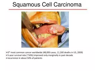
Squamous cell carcinoma (SCC) • Most commonly occurs among people with white/fair skin • Commonly located on the head, neck, forearms, and dorsal hands (sun-exposed areas) • SCC has increased associated mortality compared to basal cell carcinoma, mostly due to a higher rate of metastasis
SCC: Etiology • Cell of origin: keratinocyte • Cumulative UV exposure • Cause genetic alterations, which accumulate and provide selective growth advantage • SCC arising in nonsun-exposed areas may be related to chemical carcinogen exposure (e.g. arsenic)
SCC: Clinical manifestations • Various morphologies • Papule, plaque, or nodule • Pink, red, or skin-colored • Scale • Exophytic (grows outward) • Indurated (dermal thickening, lesion feels thick, firm) • May present as a cutaneous horn • Friable – may bleed with minimal trauma and then crust • Usually asymptomatic; may be pruritic
SCC in situ • Also known as Bowen’s disease • Circumscribed pink-to-red patch or thin plaque with scaly or rough surface • Keratinocyte atypia is confined to the epidermis and does notinvade past the dermal-epidermal junction
Back to Mr. Dominguez • Mr. Dominguez was diagnosed with invasive SCC. What is your next step in management? • Liquid nitrogen cryotherapy • Reassurance with close follow-up • Shave biopsy • Surgical removal • Topical antibiotics
Management Answer: d • What is your next step in management? • Liquid nitrogen cryotherapy (Liquid nitrogen is used to treat pre- cancerous actinic keratoses. It is NOTthe treatment for invasive squamous cell carcinoma.) • Reassurance with close follow-up (Squamous cell carcinoma is a malignant lesion with potential for metastases. You must treat it!) • Shave biopsy (You already know the diagnosis and there is no need for another biopsy.) • Surgical removal (The treatment of choice for squamous cell carcinoma is surgical excision. The specimen must be sent to pathology to document clear margins (complete excision).) • Topical antibiotics (The lesion is not an infection.)
Pathology reports for SCC • “Invasive squamous cell carcinoma” • Means there are SCC cells in the dermis • If there is no dermal involvement, it is squamous cell carcinoma in situ • Unrelated to metastatic potential • “Atypical squamous proliferation” • Often used when biopsy is too superficial • If dermis cannot be seen in the biopsy, invasive SCC cannot be excluded
SCC: Treatment • There are several medical and surgical treatment options • Suspicion of SCC should prompt referral to a dermatologist for evaluation and discussion of specific treatment approaches • Surgical Treatment Options • Surgical excision (standard of care for invasive SCCs) • Wide local excision • Mohs micrographic surgery • Curette and Desiccation (reserved for in situ SCC) • Non-surgical Treatment Options • Radiation therapy for poor surgical candidates • 5-Fluorouracil cream, imiquimod cream, photodynamic therapy – typically reserved for in situ SCCs when excision is a suboptimal choice
SCC: Course & Prognosis • For SCC arising in sun-exposed skin, the rate of metastasis to regional lymph nodes ~ 5% • Higher rates of metastasis if: • Large (diameter > 2cm), deep (> 4mm), and recurrent tumors • Tumor involvement of bone, muscle, and nerve • Location on scalp, ears, nose, and lips • Tumor arising in scars, chronic ulcers, burns, sinus tracts, or on the genitalia • Immunosuppressed patients • Tumors caused by arsenic ingestion
Patient Follow-up • All patients treated for cutaneous SCC need surveillance for the early recognition and management of: • Treatment-related complications • Local or regional recurrences • Development of new skin cancers • Patients with a history of SCC should have close follow-up • Patients are often seen every 6 to 12 months
Case Two Mr. Jenkins
Case Two: History • Mr. Jenkins is a 66-year-old man with a history of SCC who presents to the dermatology clinic for his regularly scheduled follow-up visit. He reports that during a self skin exam, he noticed a few rough, red spots on the face. He asks if this could represent another cancer. How would you describe the skin findings?
Case Two: Skin Exam Rough, scaly, thin, red-pink plaques scattered on the forehead and right temple area
Diagnosis • What is your diagnosis? Click on the correct answer. • Actinic keratosis • Basal cell carcinoma • Melanoma • Seborrheic keratosis • Squamous cell carcinoma • Verruca vulgaris
Diagnosis • That was incorrect. Try again. • Actinic keratosis • Basal cell carcinoma • Melanoma • Seborrheic keratosis • Squamous cell carcinoma • Verruca vulgaris
Your diagnosis is correct! • Actinic keratosis • Basal cell carcinoma • Melanoma • Seborrheic keratosis • Squamous cell carcinoma • Verruca vulgaris
Actinic Keratosis (AK) • AKs are premalignant lesions; they have the potential of transforming into a skin cancer. Virtually all AKs that transform into cancer will become squamous cell carcinoma (SCC). • Most AKs do not progress to invasive SCC • Risk of malignant transformation of an AK to SCC within one year is about 1 in 1000 • Risk factors for malignant progression of AK to SCC include: persistence of the AK, history of skin cancer, and immunosuppression • The keratinocyte is the cell of origin
Actinic Keratosis AKs may be considered as part of a disease spectrum:
AK: Etiology • Cumulative and prolonged UV exposure, resulting in: • UV-induced p53 tumor suppressor gene mutations • Individual risk factors can increase susceptibility: • Increasing age • Fair skin, light eyes/hair (skin types I,II) • Immunosuppression • Genetic syndromes, such as xeroderma pigmentosum and albinism
AK: Clinical manifestations • May be symptomatic (tender) • Located in sun-exposed areas • Head, neck, extensor forearms, and dorsal hands • Typically on background of sun damaged skin • Erythematous papule or thin plaque with a characteristic rough, gritty scale • Often diagnosed by feel (like sandpaper) * The diagnosis of AKs should be made cautiously in lesions > 6mm since these may represent SCC in situ or a superficial BCC.
Recognizing Sun-damaged Skin • Skin features of chronic sun damage include: • Combination of atrophy and hypertrophy • Telangiectasias • Spotty depigmentation and hyperpigmentation • Wrinkles • Skin appears “leathery” and “prematurely aged”
Solar Lentigo (lentigines) • Result from UV damage • Sun-exposed areas • One/many small brown macules
Effects of Sun Damage Cutis rhomboidalis nuchae (red neck with rhomboidal furrows) Solar Elastosis (fine nodularity, pebbly surface)
Actinic (senile) Purpura • Easy bruising • Extravasated erythrocytes and increased perivascular inflammation
Back to Actinic Keratoses
AK: Actinic cheilitis • Actinic cheilitis represents AKs on the lips, most often the lower lip • Erythematous patch with rough gritty scale involving the lower lip • Persistent ulcerations or indurated areas should prompt a biopsy to rule out malignant transformation
How would you treat this AK? • Which of the following treatments would you recommend for this AK? • Liquid nitrogen cryotherapy • Surgical excision • Radiation therapy • Topical antibiotic • Topical corticosteroids
How would you treat this AK? • Answer: a • Which of the following treatments would you recommend for this AK? • Liquid nitrogen cryotherapy • Surgical excision • Radiation therapy • Topical antibiotic • Topical corticosteroids Click here to view a video on cryotherapy
AK: Treatment • There are several topical and procedural treatment options for AKs. The best option is chosen after consideration of number, location, and thickness, among other patient factors. • Therapies are considered local – treating the individual lesion, or field therapies – treating multiple AKs in one area • Consultation with a dermatologist to guide therapy may be useful • Localized Therapies • Liquid nitrogen cryotherapy • Curettage +/- electrocautery • Shave excision • Field Therapies • Topical 5-fluorouracil or imiquimod creams • Photodynamic therapy
AK: Patient Education • Patients with AKs are at increased risk of developing other non-melanoma and melanoma skin cancers. • Therefore, these patients should have regular skin exams every 6-12 months • Patients should be seen prior to their regularly scheduled follow-up if they notice a concerning lesion on a self-skin exam
Patient Education • There are multiple resources to help educate patients about sun safety and skin cancer prevention, including: • American Academy of Dermatology: SPOT Skin Cancer™ initiative • American Cancer Society: Skin Cancer Prevention and Early Detection • The following slides are adapted from the AADSPOT Skin Cancer™program and reflect recent, Board-approved changes to public messages about sun safety: