Download
1 / 30
300 likes | 353 Views
Study evaluating TNM classification, histological malignancy scores, and prognosis correlation in tongue squamous cell carcinoma. Findings reveal TNM classification as an important prognostic indicator.

E N D
Clinical-pathological parameters in squamous cell carcinoma of the tongue Chairperson: Dr. Md. Harun-ar- Rashid Assistant Professor & Head Department of Dentistry, MMC Presenter: Dr. K M Anwar Hossain Dental Surgeon, MMCH
Authors: Dilana Duarte Lima DantasI; Carlos César Formiga RamosII; Antonio de Lisboa Lopes CostaI; • Source: Brazilian Dental Journal,. 2009 Jul 1;14 (7): E349-54. • Indexed in- Braz. Dent. J. vol.14 no.1 Ribeirão Preto June 2003
Abstract • The correlation between TNM classification and histological scores of malignancy, and the correlation of these parameters with the prognosis was evaluated in 16 cases of squamous cell carcinoma of the tongue. The cases were selected from the files of "Dr. Luiz Antônio" Cancer Hospital, Natal, RN, Brazil. After analysis of the patients' records, the data concerning TNM classification and prognosis (in a 5-year-follow-up) were obtained.
All cases were classified according to the histological malignancy grading system proposed by Anneroth et al. [Scand Dent Res 1987;95:229-249]. There was no correlation (r = 0.3083) between TNM classification and histological scores of malignancy. There was significant correlation (r = 0.7206) between TNM classification and prognosis, but there was no correlation between the histological scores of malignancy and the prognosis. It was concluded that TNM classification is an important prognostic indicator of squamous cell carcinoma of the tongue.
Introduction • Oral squamous cell carcinoma is the most frequent malignancy in the mouth, corresponding to 95% of all oral malignant lesions. The most affected sites are the tongue, inferior lips and floor of the mouth. Males are affected twice as frequently as females and the majority of patients are more than 45 years old.
When compared to other oral cancers, squamous cell carcinoma of the tongue has a great predisposition to produce metastasis in lymph nodes (incidence 15-75%) depending on the extension of the primary lesion. In clinical practice, the treatment plan and prognosis of oral squamous cell carcinoma is mainly based on the TNM classification. However, this system does not provide any information on the biological characteristics of the tumor. Information about characteristics is important to obtain new objective prognostic factors that may provide information about the aggressiveness of this kind of tumor.
The aim of this study was to assess the correlation between the TNM classification and the histological malignancy grading system proposed by Anneroth et al. and the correlation of these parameters with the prognosis of squamous cell carcinoma of the tongue. The specific objective was to assess some indicative parameters that would assist in the prognosis of these lesions and in the correct choice of therapy.
Material and Methods • Sample Selection and Morphological Analysis • Sixteen cases of squamous cell carcinoma were selected from patients' records from "Dr. Luis Antonio" Cancer Hospital (Natal, RN, Brazil). Data concerning gender, age, TNM classification and prognosis (in a 5-year-follow-up) were obtained. The TNM clinical staging was classified according to the U.I.C.C. (International Union against Cancer): stage T1 =T1N0M0; stage T2 = T2N0M0; stage T3 = T3N0M0 or T1, T2, or T3N1M0; stage T4 = any T4lesion, or any N2 or N3 lesion, or any M1 lesion.
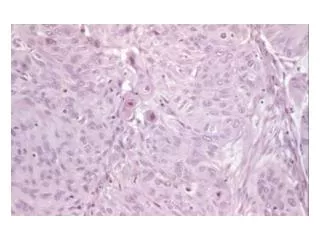
Hematoxylin and eosin (H&E) stained samples had been submitted to morphological study with optic microscopy. The histological grading system of malignancy consists of 6 morphological characteristics: degree of keratinization, nuclear polymorphism, number of mitosis, invasive pattern, "stage of invasion" and lymph plasmocytic infiltration.
As recommended by Pinto Jr. et al. the parameter "stage of invasion" was omitted because these were incisional biopsies of oral squamous cell carcinoma. All mean scores for malignancy histological grading were obtained by the sum of the scores given to each morphological parameter, and then divided by the number of used parameters. According to Anneroth et al., scores ranging from 1.0 to 2.5 are considered to be low malignancy, and those ranging from 2.6 to 4.0 are considered to be high malignancy.
Statistical Analysis • The Pearson correlation test was applied to assess the correlation between the TNM classification and malignancy histological scores and the Spearman correlation test was applied for the TNM classification and prognosis, as well as for malignancy histological scores and prognosis. The statistical tests were carried out with the GMC software, model, with a level of significance of 1%.
Results • Based on the clinical data provided by the records of patients with squamous cell carcinoma, 75% (12 cases) of the sample were males and 25% (4 cases) were females, with ages ranging from 39 to 94 years. The most frequent age group was 61 to 70 years old with 43.75% (7 patients) of all cases.
The TNM classification showed that 4 cases were classified as stage IV, 4 were stage III, 5 were stage II and 3 were stage I (Table 1). In terms of the malignancy histological scores, the results showed that 7 were classified as low score (1.2 to 2.5) and 9 were classified as high score (2.6 to 3.4) (Table 1). Results relative to the prognosis revealed that 9 patients had recurrence of the disease after the follow-up of 5 years, while 7 were free of disease.
Data analysis using Pearson correlation coefficient showed no significant statistical correlation (r = 0.3083) between the TNM classification and malignant histological scores. The Spearman correlation coefficient showed a statistically significant correlation (r = 0.7206) between TNM classification and prognosis. However, the Spearman correlation test revealed no statistically significant correlation (r = -0.1535) between the malignant histological scores and prognosis.
Discussion • With a considerably high frequency among malignant tumors of the oral cavity, squamous cell carcinoma is characterized by a disordered proliferation of cells in the squamous layer of the epithelium, where these cells express varied degrees of similarity with their cells of origin. • The endless search into histological samples of oral squamous cell carcinomas as one means of assisting in the evaluation of prognosis and in the planning of adequate therapy for the patient led the authors to conduct this study. • The data for gender (75% males, 25% females) and most frequent age group (61-70 years; 43.75%) are in agreement with Oliver et al. and Gervásio et al.
In the analysis of the variable TNM classification, the results revealed that there was a clear relation between lesion size and lymph node involvement, considering that, from the 16 cases included in this study, 8 had lesions with a diameter greater than 4 cm (lesions in stages III and IV). This fact certainly leads to a worse prognosis, if compared to lesions with smaller diameters without lymph node involvement, corroborating data from Snow and Leemans and Tytor and Oloffson. According to these authors, there is a directly proportional relation between these two factors, given that, the smaller the lesion, the easier will be the use of the conventional treatment means available for this kind of tumor, with a great possibility of cure after the first treatment.
It is important to mention that, from the 9 cases that had recurrence, 4 were in stage III, 4 were in stage IV and only 1 in stage I. This shows the great importance of the stage of the disease and its impact on metastasis. • From the 8 cases in stages I and II (Table 1), only one patient had active disease due to refusal of treatment after surgery. The remaining cases showed no recurrence of disease. This indicates that the tumor stage has an expressive significance upon its prognosis. These findings are in agreement with those reported by Snow and Leemans and Tytor and Olofsson.
When the histological malignancy grading system (1) was applied, it could also be observed that from the 7 cases classified with low scores of malignancy, 3 patients were free from the disease and 4 had recurrence. Of the 9 cases classified as high malignancy, 4 patients were free from disease and 5 had recurrent disease at a minimum follow-up of 5 years.
Based on these results, it was evident that there was no substantial correlation between the histological scores of malignancy and the prognosis, considering that 57% of the lesions classified as low scores of malignancy showed recurrence at follow-up. On the other hand, from the 9 patients classified as high malignancy, 44% of these patients were diagnosed as free from disease after the follow-up of five years. • Histological evaluation results are likely to be more precise in establishing prognosis than those essentially clinical, because histological changes can be seen microscopically before clinical changes.
From the point of view of the authors, the histological grading system proposed by Anneroth et al. modified by Pinto Jr. et al. was not an effective prognosis indicator. Therefore, the present data contradict the findings of other studies (12-17) which consider the histological grading as a valuable indicator of prognosis for oral squamous cell carcinomas. However, it is important to report that, in this study, of 9 patients with lesions with a high malignancy score, 55% had confirmed active disease at the 5-year follow-up. These data are in agreement with the above authors for considering that the higher the malignant histological grading score, the worse will be the prognosis of the lesion.
With the purpose of testing the hypothesis of correlation between TNM classification and histological malignancy grading, statistical analysis was carried out which demonstrated that there was no significant statistical correlation between the two variables. From the point of view of the authors, among the disadvantages of the histological grading system proposed by Anneroth et al. are situations in which the application of the parameters makes a large and representative biopsy sample necessary.
In addition to these aspects, Crissman et al. consider that, in histological grading, only the characteristics which demonstrate isolated prognosis value should be evaluated. A high importance should not be given to variables of difficult identification and of low isolated prognosis value, such as mitosis number and nuclear atypia, at the expense of other more objective variables and of proven significance such as tumor thickness, neural infiltration and lymphatic embolization. These authors call attention to the fact that tumors that are well differentiated are likely to invade tissues in a pattern characterized by well defined margins, while the more undifferentiated tumors infiltrate tissues in small cell groups or even as isolated cells, which may explain its correlation with the prognosis.
Based on these results, the aspects here evaluated for the prognosis and for the planning of the appropriate therapy are considered very important. It should be noted that "invasive pattern", "tumor thickness" and "inflammatory infiltration" could be important histological indicators of the degree of tumor aggressiveness, considering that they reflect the relation of the cell population with the host. In addition to this, incisional and representative biopsies should be used which provide visualization of all the invasive front and borders, as recommended by Bryne and Piffkó et al.