Download

1 / 50
500 likes | 618 Views
Unit 4 – The Integumentary System. Integumentary System. Also known as the Integument Accounts for 16% of your body weight First line of defense Almost 2m 2 Consists of 2 major components Cutaneous membrane Accessory structures. Integumentary System. Cutaneous membrane has two layers

E N D
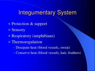
Integumentary System • Also known as the Integument • Accounts for 16% of your body weight • First line of defense • Almost 2m2 • Consists of 2 major components • Cutaneous membrane • Accessory structures
Integumentary System • Cutaneous membrane has two layers • Epidermis • Dermis • Accessory structures include: • Hair • Nails • Exocrine glands
Integumentary System • Blood vessels and nerves are found in the dermis • Deep to the dermis, is the subcutaneous layer (hypodermis)
Integumentary System • Functions of skin and subcutaneous layer: • Protection • Excretion • Maintenance of body temperature • Synthesis if vitamin D • Storage of fats • Detection of touch, pressure, pain, temperature
Integumentary System THE EPIDERMIS
The Epidermis • Stratified squamous epithelial tissue • Mostly keratinocytes (contains keratin) • Is comprised of several layers (deep to superficial) • Stratum germinativum • Stratum spinosum • Stratum granulosum • Stratum lucidum • Stratum corneum • Thin Skin – covers most of your body • Thick skin – palms of hands, soles of feet
Epidermal Layers • Stratum germinativum • Deepest layer • Attached to basal lamina • Has epidermal ridges • Extend into dermis to strengthen bond between epidermis and dermis • Pattern of ridges are revealed in fingerprints • Dominated by basal cells (germinative) • Stem cells that replace lost superficial cells • Contains melanocytes – cells that make melanin
Epidermal Layers • Stratum spinosum • 8-10 layers of cells • Made of freshly divided basal cells
Epidermal Layers • Stratum granulosum • “grainy layer” • 3-5 layers of cells pushed superficially from stratum spinosum • Make large amounts of keratin • Basis of hair and nails in humans • Makes skin waterproof • Cells die in this layer
Epidermal Layers • Stratum lucidum • Found in the palms of your hands and soles of your feet
Epidermal Layers • Stratum corneum • The exposed surface of the skin • 15-30 layers of keratinized cells • It takes 15-30 days for cells to move from the stratum germinativum to the stratum corneum • Cells spend about 2 weeks in the stratum corneum before being shed
Epidermal Layers • Stratum corneum (con’t) • Is water resistant, but not waterproof • Water evaporates at a rate of about 500ml/day • Called insensible perspiration • Sweat glands produce sensible perspiration • Blisters are formed when the connections between superficial and deeper layers are damaged. • Fluid collects between layers
Integumentary System THE DERMIS
The Dermis • Lies between the epidermis and the subcutaneous layer • Two major components • Superficial papillary layer • Deeper reticular layer
The Papillary Layer • Superficial portion of dermis • Areolar tissue • Contains capillaries, lymphatics, and sensory neurons that supply the surface of the skin • Contain dermal papillae that interlock with the epidermal ridges
Reticular Layer • Deeper portion of dermis • Dense irregular connective tissue • Collagen and elastic fibers • Contains all cells of connective tissue proper • Also contains blood vessels, lymph vessels and nerve fibers
Dermal Strength and Elasticity • Dermis contains both collagen and elastic fiber • Allows dermis to tolerate limited stretching • Water also helps with flexibility and resilience • Pinch test for dehydration • Aging, hormones, UV radiation damages elastic fibers • Leads to wrinkles and sagging skin
Dermal Strength and Elasticity • Extensive stretching of dermis can exceed the elastic capabilities of the skin • Complete recoiling of fibers is prevented • Result is stretch marks • Retin-A (derivitave of Vitamin A) can help repair the dermis and can lessen appearance of wrinkles and stretch marks.
Lines of Cleavage • The collagen and elastic fibers in the dermis are usually arranged in parallel bundles • Arranged to resist forces during normal movement • The pattern forms lines of cleavage
Lines of Cleavage • Why are the lines important? • Cuts parallel to the lines of cleavage will remain closed and heal with minimal scarring • Cuts perpendicular to the lines will be pulled open by the recoiling elastic fibers and will result in greater scarring.
Integumentary System THE SUBCUTANEOUS LAYER
The Subcutaneous Layer • Also known as the hypodermis • Consists of areolar and adipose tissue • Very elastic • Superficial region contains blood vessels • Subcutaneous fat serves as an insulator and a major energy reserve for the body.
The Subcutaneous Layer • At puberty, distribution of fat starts to differ between the sexes • Men tend to store fat at the neck, arms, low back, and buttocks • Women tend to store fat at the breasts, buttocks, hips and thighs • Both can accumulate fat in the abdominal region
Integumentary System Accessory structures
Hair and Hair Follicles • Hair – cover almost every surface of your skin • Exceptions are sides and soles of feet, palms of hands, sides of toes and fingers, lips, portions of external genitalia • Body has 2.5 million hairs • 75% NOT on your head • Hair is a nonliving structure produced by hair follicles • Made of the protein keratin
Hair and Hair Follicles • Function of hair on head • Protect from UV radiation • Cushion light blows to head • Insulate skull • In nostrils and ear: • Prevent entry of foreign particles • All hair serve as sensory receptors
Hair and Hair Follicles • Have arrector pili muscles attached to hair follicle • When contracted, makes hair stand up • From emotions, cold (goose bumps) • In furry mammals, this will increase insulation, but doesn’t have that effect in humans
Hair and Hair Follicles • Hair will grow for 2-5 years before being shed • Grows about 0.33mm each day • While growing, the follicle will incorporate nutrients/chemicals into the hair shaft • Can be useful for detecting disorders or drugs. • Can be used for DNA fingerprinting if hair contains nucleated cells
Hair and Hair Follicles • Hair coloration is from the melanocytes producing varying amounts of melanin in the hair follicle • Different forms of melanin will give hair a dark brown, yellow-brown, or red appearance
Glands In the Skin • Sebaceous (Oil) Glands • Can secrete oil from hair follicle or directly onto skin • The secreted material is called sebum • Made of fatty acids, cholesterol, proteins, and electrolytes • Inhibits bacteria growth, lubricates and protects hair, and conditions surrounding skin
Glands In the Skin • Sebaceous (Oil) Glands (Con’t) • Sebaceous follicles • Glands NOT associated with hair follicles • Discharge sebum directly onto epidermis • Located on face, back, chest, nipples, external genitalia • Very active just before birth and during puberty
Glands In the Skin • Sweat Glands • Also known as sudoriferous glands • Two types • Apocrine sweat glands • Found in armpits, around nipples, and pubic region • Secrete sweat into hair follicles • Sticky, cloudy, odorous secretion • Begin secreting at puberty • This sweat is a nutrition source for bacteria, which intensifies odor
Glands In the Skin • Sweat Glands (Con’t) • Two types • Merocrine sweat glands • Discharge secretion directly onto surface of skin • More numerous (2-5 million) than apocrine glands • Palms and soles have highest number • 3000 per square inch • Secretion is 99% water with some electrolytes (mainly sodium chloride)
Glands In the Skin • Sweat Glands (Con’t) • Two types • Merocrine sweat glands • Functions: • Cool surface of skin (maintain homeostasis) • Excrete water and electrolytes • Provide protection • Dilute harmful chemicals • Discourages growth of mmicroorganisms
Nails • Protect exposed dorsal surfaces of the tips of the fingers and toes • Parts: • Nail body – visible portion of nail • Nail bed – lies under nail body • Lateral nail grooves – lateral borders of nail body • Lateral nail folds – portion of skin overlapping lateral portion of nail body • Free edge – distal portion extending past nail bed
Nails • Parts (con’t): • Hyponychium – thickened skin at distal end of nail bed • Nail root – forms the nail • Eponychium (cuticle) – skin flap covering proximal nail body • Lunula – pale crescent at proximal end of nail body
Integumentary System Injury Response
Injury Response • Excellent regeneration capabilities and response to stresses. • Calluses form when repeated stresses from manual labor are placed on the skin • The stem cells of stratum germinativum divide more rapidly, thickening the skin in that area
Injury Response • The process of injury repair can be slow • Infection and fluid loss can complicate repair • The type of injury also determines rate of repair • Thin, straight cut (incision) will generally heal more quickly than a scrape (abrasion) because of the relative surfaces involved
Injury Response • Bleeding occurs when the damage extends into the dermis. Why? • No vessels in epidermis
Injury Response • Steps to injury response Step 1 • Bleeding occurs and mast cells trigger the inflammatory response
Injury Response Step 2 • Blood clot (scab) forms • Restores integrity of epidermis • Restricts microorganisms from entering area • Stratum germinativum cells migrate along edges of wound • Divide rapidly to replace epidermal cells • Macrophages patrol area collecting debris and pathogens
Injury Response Step 2 (con’t) • Increased capillary formation enhances blood flow • Combination of blood clot, fibroblasts, and new capillary network is called granulation tissue
Injury Response Step 3 • Over time, deeper portion of clot dissolves • Number of capillaries decline • Fibroblast activity leads to increase in collagen fibers and ground substance • Epidermal cells have migrated over the network of collagen fibers
Injury Response Step 4 • After several weeks, the scab is shed • The repairs do not restore the integument to its original condition • Dermis will contain abnormally large numbers of collagen fibers, and few capillaries • Severely damaged hair follicles, glands, muscle and nerve cells are seldom repaired • Replaced by fibrous tissue (Scar)
Integumentary System Aging
Aging and the Integument • Effects of aging on the integument • Thinning epidermis • Decreased vitamin D production • Muscle and bone weakness • Decreased melanocyte activity • More sensitive to sun • Decline in glandular activity • Dry, scaly skin. Less perspiration means increased overheating • Reduced blood supply to dermis
Aging and the Integument • Effects of aging on the integument • Hair follicles stop functioning • Thinner, finer hair • Dermis thins • Sagging and wrinkling occur • Decrease in sex hormones • Hair characteristics and fat distribution change • People of both sexes age 90-100 tend to look alike • Skin repairs more slowly • Repairs can take twice as long as a young adult • Recurring infections may result