Download
1 / 1
10 likes | 120 Views
SPIO Nanoparticle Labeling of Rat Microglia Cells Utilizing MR Microscopy Michelle A. Sokoll 1,2 , Jens T. Rosenberg 1,2 , Zaki Estephan 3 , Megan Muroski 3 and Samuel C. Grant 1,2

E N D
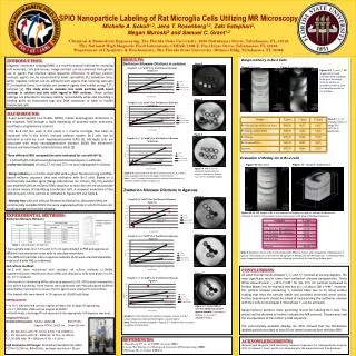
SPIO Nanoparticle Labeling of Rat Microglia Cells Utilizing MR Microscopy Michelle A. Sokoll1,2, Jens T. Rosenberg1,2, Zaki Estephan3, Megan Muroski3 and Samuel C. Grant1,2 1Chemical & Biomedical Engineering, The Florida State University, 2525 Pottsdamer Street, Tallahassee, FL, 32310. 2The National High Magnetic Field Laboratory, CIMAR, 1800 E. Paul Dirac Drive, Tallahassee, FL 32310. 3Department of Chemistry & Biochemistry, The Florida State University, Dittmer Bldg, Tallahassee, FL 32306. INTRODUCTION: Magnetic resonance imaging (MRI) is a multi-functional method for analyzing soft materials, cells and tissues. Image contrast can be enhanced through the use of agents that improve signal relaxation efficiency. To achieve positive contrast, agents can be constructed to lower spin-lattice (T1) relaxation times while negative contrast can be achieved with agents that lowering spin-spin (T2) relaxation times. Iron oxides are common agents that exhibit strong T1/T2contrast [1]. This study aims to evaluate iron oxide particles with novel coatings in solution and cells with regard to MRI contrast. These surface coatings are intended to increase stability and solubility while also providing a binding point for fluorescent tags and DNA sequences to label or modify transfected cells. RESULTS: ZwitterionSiloxane Dilutions in solution Bangs-mCherry in Bv-2 Cells Figure 2 Figure 8: Spin echo Figure 9: Gradient recalled echo Figures 8-9. T2 and T2* MR images of Bv-2 cells incubated with a stepwise build up of the Bangs-mCherry particle as indicated in Table 8, with the complete particle in the top layer. Figure 3 • BACKGROUND: • Super-paramagnetic Iron Oxides (SPIOs) induce paramagnetic distortions in the magnetic field through a rapid dephasing of proximal water molecules, resulting in a hypointense contrast. • The Bv-2 cell line used in this study is a murine microglia that plays an important role in the brain’s immune defense system. Bv-2 cells can be activated in vitro by e-coli lipopolysaccharide (LPS) [2]. Microglia cells are associated with many neurodegenerative diseases (NDD) like Alzheimer's disease and Amyotrophic Lateral Sclerosis (ALS) [3]. • Three different SPIO nanoparticles were evaluated for use with BV-2s: • 3-(dimethyl(3-(trimethoxysilyl)propyl)ammonio)propane-1-sulfonate, (zwitterion siloxane) of sizes 2.7 nm and 17.5 nm was investigated in solution. • Bangs-mCherry is a micron-sized SPIO with a green fluorescent tag and DNA-based mCherry sequence that was evaluated with Bv-2 cells. Based on a commercially available agent (Bangs Laboratories Inc, Fishers, IN), this particle was modified with an mCherry DNA sequence to stain the cell red and provide an optical means of identifying transfected cells. A stepwise evaluation of the different parts of the particle as indicated in Figures 8-9 and Table 8. • Molday Iron with and without Rhodamine (BioPal Inc ,Worcester/MA) are commercially available SPIOs that were evaluated with Bv-2 cells for future cell tracking usage and bimodal imaging. Table 8. T1,T2,T2* relaxation times of Bangs SPIO particles in agarose gel. Figure 4 Evaluation of Molday Ion in Bv-2 cells Figure 10: Spin echo Figure 11: Gradient recalled echo Figure 2-4. MRI of 2.7 nm zwitterionsiloxane dilution samples. From top to bottom: T1, T2 and T2* weighted images. Graph 2-4. Concentration of zwitterionsiloxaneverses R1, R2 and R2*. Linear relaxivity is observed with higher concentrations. Note: T1 and T2 contrast increased with increased concentrations of zwitterion siloxane shown in Figures 2-4. Zwitterion Siloxane Dilutions in Agarose Figure 5 Figures 10-11. MR images of Bv-2 cells exposed to Molday Iron with or without Rhodamine as indicated in Table 9. The greatest contrast can be seen with 60mg of MoldayRhodamine. EXPERIMENTAL METHODS: Zwitterion Siloxane Dilutions Figure 1. Diagram of solutions zwitterion siloxane Figure 6 Table 1. Concentrations of Zwit Iron • Two sample sizes of 2.7 nm and 17.5 nm were diluted in PBS and agarose at different concentrations to be able to calculate relaxivities. • The different particles sizes in agarose samples (A-G) were scanned separately in pairs of 8 with PBS as reference. Table 9. Relaxation times of Bv-2 cells labeled with different masses and conjugations of Molday Ions in agarose. The shortest T2 is seen for the 60-mg layer of Molday Ion with Rohodamine. T2 relaxation show some irregularity likely due to inconsistent layering, particularly for the 60-mg Molday layer. • Cell culture methods • Bv-2 cells were maintained with standard cell culture methods (a-DMEM supplemented with Fetal Bovine Serum (FBS) and antibiotics). Cells were grown in a 5% CO2, 37 C incubator. • 24 hrs prior to introducing SPIOs, cells were exposed to LPS. SPIOs were incubated for 6 hrs before harvesting. Three washes were performed with PBS (phosphate-buffered saline) before trypsination to ensure that no agents were attached to cell surfaces. • The labeled cells were layered in 1% agarose at 150,000 cells/layer. CONCLUSIONS: All experimental results showed T1, T2 and T2* contrast at varying degrees. The most significant results came from zwitterion siloxane nanoparticles. These SPIOs showed lower r2 (19.9 s-1mM-1 for the 17.5 nm particle) compared to Feridex (Bayer, Inc) for example that has an r2 of about 160 s-1mM-1. However, this particle showed interesting T1 contrast likely due to its novel surface coating that kept the particle stable and provided enhanced water access. Further experiments should be aimed at incorporating the zwitterion siloxane with Bv-2 cells to investigate if intracellular T1 can be achieved. Bangs-mCherry particles show promising results for labeling Bv-2 cells. This project will be directed to further evaluate the MR contrast, fluorescence and the incorporation of the mCherry DNA sequence. The commercially available Molday ion SPIO showed that the Rhodamine labeled particle provided at least (if not better) contrast than the bare SPIO. Figure 7 • MR Equipment: • 11.75-T, 500-MHz WB vertical magnet at FAMU-FSU College of Engineering • 21.1-T, 900-MHz UWB vertical magnet at NHMFL • At both fields, a birdcage RF coil resonant at the appropriate 1H frequency was used. • Imaging Protocols: • Standard Parameters: Matrix=128x128 BW=75 kHz • Approx. FOV=1.5x1.0 cm Slice=1.0 mm • T1 : 2D Spin Echo with TE= 10 ms; 8 TRs = 50-15000 ms • T2 : 2D Spin Echo with TR = 5000 ms; 15 TEs = 8-128 ms • T2*:2D GRE with TR = 5000 ms; 8 TEs = 5-35 ms • High Resolution Cell Images: 3D Gradient Recalled Echo (GRE): • TE/TR=7.5/150 ms, BW=55 kHz, Isotropic resolution = 50 μm Figure 5-7. Proton MRI of 17.5-nm zwitterion siloxane agarose samples. From top to bottom: T1, T2 and T2* weighted images. Graph 5-7. Concentration of zwitterionsiloxanevs R1,R2 and R2*. Linear relaxivity is observed with higher concentrations. Note. Similar contrast displayed in Figures 5-7. Similar values are seen in agarose and solutions. REFERENCES: 1.Rosenberg JT. et. al CMMI In press2011 2. Long Tai Zhenget al European Journal of Pharmacology 2008. 3.McGeer PL et al Glia1993 (7). ACKNOWLEDGEMENTS: National High Magnetic Field Laboratory: Research Experience for Undergraduates Program 2011. Dr. Samuel C. Grant and Dr. Jens Rosenberg for the opportunity and their guidance.