Download
1 / 75
770 likes | 1.21k Views
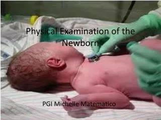
0. The Newborn Examination. Karen Montgomery-Reagan, D.O. OUCOM Department of Pediatrics. Overview. Review birth Hx and VS Listen to heart while infant quiet: Cardiovascular- inspection, palpation, and auscultation

E N D
0 The Newborn Examination Karen Montgomery-Reagan, D.O. OUCOM Department of Pediatrics
Overview • Review birth Hx and VS • Listen to heart while infant quiet: Cardiovascular- inspection, palpation, and auscultation • Start from top: Head and Neck – head, face, ears, eyes, nose, mouth, and neck • Chest and Lungs • Abdomen – inspection, palpation, and auscultation • Genitourinary – kidneys and genitalia • Orthopedic – spine, upper and lower extremities
Vital Signs • Heart Rate (rates/min) • Awake 85-205 • Sleeping 80-160 • Respiratory Rate (breaths/min) • 30-60 • Systolic Blood Pressure (SBP) • Infant SBP: 80 • Age 12 hours: 50-70 / 25-45 • Age 96 hours: 60-90 / 20-60 • Hypotension defined in neonate to 28 days <60
0 APGAR Score
External Characteristics To be done within few hrs of birth Edema Skin texture, color, and opacity Lanugo Plantar creases Nipples and breasts Ear form and firmness Genitals Neuromuscular Score To be done within 24 hrs Posture Square Window Arm recoil Popliteal angle Scarf sign Heel to ear Dubowitz/Ballard Score
0 Small for Gestational Age • Symmetric • HC, length, weight all <10 percentile • 33% of SGA infants • Cause: Infection, chromosomal abnormalities, inborn errors of metabolism, smoking, drugs • Asymmetric • Weight <10 percentile, HC and length normal • 55% of SGA infants • Cause: Uteroplacental insufficiency, Chronic hypertension or disease, Preeclampsia, Hemoglobinopathies, altitude, Placental infarcts or chronic abruption • Combined • Symmetric or asymmetric • 12% of SGA infants • Cause: Smoking, drugs, Placental infarcts or chronic abruption, velamentous insertion, circumvallate placenta, multiple gestation
0 Large for Gestational Age • Etiologies • Infants of diabetic mothers • Beckwith-Wiedemann Syndrome • characterized by macroglossia, visceromegaly, macrosomia, umbilical hernia or omphalocele, and neonatal hypoglycemia • Hydrops fetalis • Large mother
0 Skin • Color • Pallor: associated with low hemoglobin • Cyanosis: associated with hypoxemia • Plethora: associated with polycythemia • Jaundice: elevated bilirubin • Slate grey color: associated with methemoglobinemia
0 Skin • Jaundice • Total and direct bilirubin levels should be measured in newborns with jaundice. • Causes can be related to increased unconjugated hyperbilirubinemia (physiologic, breastfeeding, increased production, decreased hepatic uptake/conjugation) or conjugated (hepatobiliary disorders, ductal disturbances) • The AAP has published guidelines on the management of hyperbilirubinemia in the newborn.
0 Skin • Lanugo • Fine hair on shoulders and back • More common in preemies • Usually disappears in 2-4 weeks • Vernix Caseosa • Cheesy white covering
0 Skin Lesions • Petechiae • On scalp and face after vertex delivery • Sucking blister • Develops from infant sucking on skin in utero. • Disappears as infant progress with feeding. • Hemangiomas • Usually enlarges in 1st year of life. • Large facial hemangiomas may be associated with posterior fossa malformations. • MC benign tumor of infancy • Most require no intervention
0 Skin Lesions • Telangiectatic Nevi • Also called salmon patches or stork bite nevi • Flat, pink lesion found on upper eyelids, forehead, nape of neck • Fade by 1-2 years old except those on nape of neck which may persist • Cutis Marmorata • Transient mottling of the skin • Occurs when baby is exposed to the cold
0 Skin Lesions • Milia • Pinpoint white papules of keratogenous material. • Usually on nose, cheeks and forehead. • Can last for several weeks.
0 Skin Lesions • Miliaria • Obstructed eccrine sweat ducts. • Pinpoint vesicles on forehead, scalp, and skinfolds. • Usually clears within one week.
0 Skin Lesions • Transient Neonatal Pustular Melanosis • Small vesicopustules, generally present at birth. • Contain WBCs and no organisms. • Intact vesicle ruptures to reveal a pigmented macule surrounded by a thin skin ring.
0 Skin Lesions • Erythema Toxicum Neonatorum • Most common newborn rash. • Lesions are firm, yellow or white pustules on a red and swollen base. • Lesions may be found in face, trunk and limbs. • Lasts about 5-7 days. • Wright Stain shows eosinophil sheets.
0 Skin Lesions • Café au lait spots • Suspect nuerofibromatosis if there are many large spots
0 Skin Lesions • Mongolian spots • Well demarcated symmetric bluish gray to deep brown to black skin markings • Common among people of Asian, East Indian, African, and Latino heritage. • Often on the base of the spine, on the buttocks and back • Generally fade in a few years and disappear by puberty.
Neurological Exam • Posture • Term infants normal posture is hips abducted and partially flexed, with knees flexed. • Arms are abducted and flexed at the elbow. • Fists are often clenched, with the fingers covering the thumb • Tone • To test, support the infant with one hand under the chest. Neck extensors should be able to hold head in line for 3 seconds • There should be no more than 10% head lag when moving from supine to sitting positions.
0 Neurological Reflexes • Ensure symmetrical • Biceps jerk C5-C6, knee jerk L2-L4, ankle jerk S1-S2, anal wink S4-S5, truncal incurvation reflex T2-S1. • Primitive reflexes are present: Babinski, Moro, Galant, palmer and plantar grasps, sucking, rooting, placing/stepping, and asymmetric tonic neck reflex (Fencer’s pose).
0 Neurological Exam • Cranial Nerve Exam • CN II: Pupillary reflex • Oculocephalic Reflex (doll’s eyes) • CN III, IV, and VI • If Brainstem intact: • Eyes deviate contralaterally • Look away from direction of rotation • CNV and VII: Corneal, sucking, and rooting reflexes • CN VIII: Response to Noise • CN IX and X : Gag reflex
0 Macrocephaly • Causes • Familial with autosomal dominant inheritance • Hydrocephalus • Other conditions • Achondroplasia (skeletal dysplasia) • Sotos' Syndrome (Cerebral Gigantism) • Alexander's Disease • Canavan's Disease • Gangliosidoses • Glutaric aciduria Type I • Neurofibromatosis Type I
0 Microcephaly • Causes • Familial • Trisomy 13, 18, 21 • Cornelia de Lange, Rubinstein-Taybi, Smith-Lemli-Opitz, Prader-Willi • Teratogen Exposure • Fetal Alcohol Syndrome • Radiation exposure in utero (<15 weeks gestation) • Fetal Hydantoin • TORCH Virus congenital infection • Cytomegalovirus, Rubella, Toxoplasmosis • Other causes • Meningitis/Encephalitis • Gestational Diabetes • Maternal hyperphenylalaninemia • Hypoxic-ischemic encephalopathy
0 Head Examination • Head • Check overriding sutures, number and size of fontanelles • Molding • Over-riding of cranial bones • Normal finding at delivery • Resolves spontaneously over first 5 days of life
0 Head Trauma • Cephalohematoma • Not as common, but can occur after prolonged labor and instrumentation use. • Rupture of blood vessels that traverse skull to the periosteum • Fluctuant swelling, does not cross suture lines • No overlying discoloration, but possible fracture • Uncomplicated resolves in 2 weeks to 3 months, if fracture, Xray in 4-6 weeks to ensure closure, depressed fractures require neurosurgical consult.
0 Head Trauma • Caput seccedaneum • Common after prolonged labor. • Accumulation of blood above periosteum. • Soft tissue swelling that crosses suture lines with overlying petechiae, purpura or bruising. • Usually resolves spontaneously over several days.
Anterior Junction of coronal suture and sagittal suture Mean newborn size: 2.1 cm (larger in black infants) Often enlarges in first few months of life Closes between 4 to 26 months (median 13.8 months) Closes by 3 months in 1% of infants Closes by 24 months in 96% of infants Posterior Junction of lambdoidal suture and sagittal suture Mean newborn size: 0.5 to 0.7 cm Closes by 2 months Fontanelles
0 Fontanelles • Exam of anterior fontanelle • Palpate fontanelle with infant sitting upright quietly • Auscultate for bruit (suggests AV malformation) • Macewen's Sign (percussion of fontanelle) • Dull cracked-pot sound suggests increased ICP
0 Fontanelles • Bulging fontanelle • Crying, coughing or vomiting • Increased intracranial pressure: Hydrocephalus, Meningitis/encephalitis, Hypoxic-ischemic injury, Intracranial hemorrhage, Dermoid tumors of the scalp • Sunken fontanelle • Decreasedintracranial pressure (dehydration) • Large fontanelle or delayed closure • Congenital hypothyroidism,Trisomy 21, Rickets, Achondroplasia, Increased Intracranial Pressure
0 Head Examination • Craniosynostosis • Premature closing of cranial sutures • Results in growth restriction perpendicular to affected suture and compensatory overgrowth in unrestricted regions. • If accompanied by restricted brain growth or hydrocephalus, neurosurgical intervention is needed.
0 Facial Examination • Facial Nerve Paralysis • Usually caused by compression of the facial nerve against the sacral promontory or by trauma of a forceps delivery. • The nasolabial fold on the affected side is not present, the corner of the mouth droops and the affected eye is unable to close. • Infant will have difficulty with feeding, drooling from the paralyzed side. • Most palsies resolved spontaneously within days.
0 Ear Examination Assess for asymmetry or irregular shape • Note presence of auricular or pre-auricular pits, fleshy appendages, lipomas, or skin tags. • Low set ears • Below lateral canthus of eye • Associated with genitourinary anomalies, because these areas develop at similar times. • Malformed ears • Can be associated with Downs or Turners Syndromes
0 Eye Examination • Normal Eye findings following delivery • Red reflex • Hold opthalmoscope 6-8” from eye. • Should transmit a clear red color back. • Equal pupil size and reactivity to light • Retinal or Subconjunctival Hemorrhages • Common after vaginal delivery • Clears spontaneously in 1-2 weeks • Lid edema • Force applied to open the eye often results in lid eversion • Examination should be postponed until the edema resolves • Eye Color • Permanent eye color usually not attained until age 6 months.
0 Eye Examination • Conjunctivitis • Relatively common in newborns. • Chemical • Silver nitrate prophylaxis given at delivery • Requires no treatment, • resolves within 48 hours • Gonorrheal • 24-48 hours old • Profound edema, purulent exudate • Tx – Penicillin G, Rocephin, or Claforan • Chlamydial • Occurs within 7-10 days • Watery discharge changes to copious/purulent • Tx – Erythromycin • HSV • Occurs within 2 weeks • Keratitis, cataracts, chorioretinitis • Tx – Topical and systemic antivirals
0 Eye Examination • Abnormal Funduscopic Exam • Lens opacity • Indicates congenital cataract • Associated with TORCH Virus infection • If monocular or dense cataract, newborn is at risk for developing amblyopia. • Leukocoria (White reflex or Cat’s eye reflex) • Suggests lens, vitreous or fundus abnormality • Evaluate for Retinoblastoma • Requires Opthalmologic Referral
0 Eye Examination • Coloboma (ocular tissue defect) • Eyelid margin defect: Treacher Collins Syndrome • Aniridia (absent iris) • Usually occurs bilaterally • Associated with poor visual acuity and nystagmus • Iris and retina defect: CHARGE association • Coloboma • Heart disease • Choanal Atresia • Postnatal growth retardation • Genital hypoplasia • Ear Abnormality • Requires Opthalmologic Referral
0 Nose Examination • Babies are obligate nose breathers until 4 months old. • Check patency with stethoscope (listen over nares). • Look for nasal flaring as a sign of increased respiratory effort. • Choanal Atresia • Small NG tube unable to pass through nares. • Normally should meet no resistance. • Bilateral atresia • Cyanosis that is relieved with crying. • Is an emergency in the newborn. • Requires an oral airway and surgical repair
0 Mouth Examination • Observe the size and shape of the mouth • Microstomia – seen in Trisomy 18 and 21 • Macrostomia – seen in mucopolysaccharidoses • Fish mouth – seen in Fetal Alcohol Syndrome • Macroglossia – associated with hypothyroidism and mucopolysaccharidoses • Small Chin – associated with Pierre Robin
Epstein pearls small white cysts which contain keratin frequently found on either side of the median raphe of the palate. Resolves in 1-2 months Natal Teeth Occurs 1 in 2,000 births Location is most often the lower incisors. Risk of aspiration if loosely attached, most require removal. More often present in newborns with cleft palate. Mouth Examination
0 Mouth • Ranulas • small bluish-white swellings of variable size on the floor of the mouth representing benign mucous gland retention cysts
0 Mouth • Cleft Lip • Incidence 1 in 750 (Caucasian) • Can be seen with or without cleft palate • Common in Trisomy 13 • Repair is usually at 3 months of age • Cleft Palate • Incidence 1 in 2500 (Caucasian) • Midline defect starts at uvula • May involve soft and hard palate and incisive foramen • Repair is usually before age 1 for normal speech
0 Neck Examination • Palpation • Palpate all neck muscles • Webbed neck • Associated with Turner’s and Noonan’s Syndromes • Lymph Nodes • Unusual at birth, presence usually indicates congenital infection • Torticollis • Sternocleidomastoid muscle injury from birth trauma. • Hematoma and fibrosis results in muscle shortening. • Muscle adaptation from abnormal intrauterine position.
0 Neck Examination • Most common neck masses: • vascular malformations, abnormal lymphatic tissue, teratomas, and dermoid cysts • Cystic hygromas • Most common neck mass • Thyroglossal duct cysts • Typically midline and inferior to hyoid bone • Surgical consultation is required
0 Chest/Lung Examination • Inspection • Supernumerary breast or nipple is common (10%) • Breast enlargement secondary to maternal hormones • Unilateral absence or hypoplasia of pectoralis major • Poland's Syndrome (Poland's Sequence) • Widely spaced nipples • Turner's Syndrome • Noonan Syndrome
0 Chest/Lung Examination • Inspection • Chest Deformity • Pectus Carinatum • Much less common than Pectus Excavatum • More common in males by ratio of 4:1 • Narrow thorax with increased anteroposterior diameter • Pectus Excavatum • Gender predominance: Boys (3:1 ratio) • Mild: Oval pit near infrasternal notch • Severe: Sinking of entire lower sternum
0 Chest/Lungs • Observe • Respiratory pattern • Brief periods apnea are normal in transition, called “periodic breathing” • Chest movement • Symmetry • Retractions and Tracheal tugging • Ascultation • Audible stridor, grunting • Wheeze, rales, rhonchi
0 Cardiovascular • Inspection • Check for pallor, cyanosis, or plethora • Palpation • Check capillary refill • Should be less than 3 seconds • Precordium • Feel for increased activity and thrill • Pulses • Ipsilateral femoral (postductal) and brachial (preductal) pulses should be equal in timing and intensity • Decreased femoral = coarctation of aorta • Bounding pulses often indicated PDA
0 Cardiovascular • Auscultation • Rhythm • Gallup Rhythms • Split S2 is Normal • Split S2 absent • Aortic or pulmonic valvular atresia or stenosis • Results in high pulmonary vascular resistance • Rate • Regular, irregular • Murmur • Quality, radiation, location of intensity
0 Abdominal Inspection • Abdominal Shape • Flat: Seen with decreased tone, Diaphragmatic hernia - abdominal contents in chest, Abnormal abdominal musculature • Distended: Consider obstruction, Excess air inside or outside the bowel, Fluid in the peritoneal cavity (ascites), Enlarged viscus