Download
1 / 81
810 likes | 986 Views
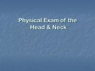
Physical Exam of the Head & Neck. INTRODUCTION. It is usually the initial part of a general physical exam, after the vital signs.

E N D
INTRODUCTION • It is usually the initial part of a general physical exam, after the vital signs. • It begins with inspection, and then proceeds to palpation. It requires the use of several special instruments in order to inspect the eyes and ears, and special techniques to assess their special sensory function.
EXAMINATION of the HEAD Inspection • Observe the patient's facial expression and appearance. Look for symmetry, size, shape, masses and involuntary movements • Hair: Observe quantity, distribution, texture, pattern of any hair loss. Look for lice or nits (the eggs of lice) • Scalp: Part the hair in several places and look for scaliness, erythema, skin lesions and nodules
Palpation • Palpate with finger pads • Generally, palpation is done only if patient symptomatic (head pain, trauma, etc.) • Skull: deformity from trauma, muscular tenderness from tension headaches • Temporal arteries: thickening, tenderness, or absent pulse in temporal arteritis • Hair: texture may change in thyroid disease, becoming more coarse • Palpation of the lymph nodes
Deformities of skull (cranium) • Microcephaly a congenitally small skull resulting from failure of the brain • Macrocephalus is an abnormally large head due to hydrocephalus, Paget’s disease (osteitis deformans), and acromegaly • Oxycephaly (steeple skull) characterized by a long anteroposterior axis, narrow in width, and pointed at the vertex. It is caused by premature union of the cranial sutures
Apert syndrome a form of acrocephalosyndactyly steeple skull + syndactyly
ANATOMY OF THE EAR • External ear • Auricle (or pinna) and external auditory canal are cartilage covered with thin, sensitive skin • Cerumen secreted from distal 1/3 of canal- protects skin • Middle ear: • Tympanic membrane (TM) normally looks "pearly gray" • Pars tensa- inferior 2/3 • Pars flaccida- superior 1/3 (covers the chorda tympani) • Umbo- where malleus attaches to TM, • Malleus- manubrium (handle) and short process are visible • Eustachian tube- equalizes middle ear pressure • Inner ear: • The cochlea and semicircular canals
EXAMINATION of the EAR • Inspection • External ear - observe position and shape, inspect for symmetry, lesions, drainage from external auditory meatus • Position: Top of auricle should be above line drawn between outer canthus of eye and occipital protuberance. Low set auricle may signify chromosomal abnormality. • Possible findings • Tophi- deposits of uric acid crystals found in patients with gout • Chondritis- infection of cartilage, often caused by piercing • "Cauliflower"-repeated trauma causes cartilage necrosis • Otitis externa- "swimmer's ear", pulling on lobe often painful • Skin cancer - often nodular, with induration, scaling and superficial ulceration.
Middle ear - otoscopic exam • Insert otoscope slowly, avoiding bumping the canal • Cerumen removal may be necessary • Cerumen spoon- often causes EAC bleeding • Irrigation - contraindicated if TM perforation • Removal with direct visualization • Pneumatic otoscopy • assesses mobility and compliance of TM • Effusion (fluid in middle ear) will hamper TM mobility • Retraction from eustachian tube dysfunction may allow movement only with negative pressure • Findings • Mobility • Bulging, no mobility Pus in middle ear- otitis media (OM) • Retracted, no mobility Eustacian tube dysfunction +/- effusion • Color • Red Infection, crying • Deep red or blue Blood (from trauma) • White flecks, plaques Healed inflammation • Bubbles Serous fluid
The Ear-Hearing assessment • Response to questions during history • Response to a whispered voice • Tuning fork air/bone conduction • Rinne (left) • Weber (right)
ANATOMY OF THE EYE • External eye: Eyelids, lacrimal gland and duct, palpebral fissures, medial and lateral angles. • Internal eye: Light travels through cornea, anterior chamber, pupil, lens, and vitreous body on the way to the retina. • Fundus: The posterior structures of the eye include the retina, retinal arteries and veins, the optic disc and the macula. These structures are viewed with the ophthalmoscope
ANATOMY OF THE EYE External eye: Eyelids, lacrimal gland and duct, palpebral fissures, medial and lateral angles. Internal eye: light travels through cornea, anterior chamber, pupil, lens, and vitreous body on the way to the retina. Fundus: The posterior structures of the eye include the retina, retinal arteries and veins, the optic disc and the macula. These structures are viewed with the ophthalmoscope
EXAMINATION of the EYE • Vision testing • Should be done with any visit involving an eye complaint • Used to screen children for visual problems • Acuity: • Far vision - test at 6 m with Snellen chart • Patient covers one eye, and is instructed to read the smallest line possible. Patient must correctly read half of symbols on line. Repeat for other eye. • Near vision - test at 35cm with pocket chart • Patient covers one eye, and is instructed to read smallest line possible. Repeat for other eye. • Visual fields: • Confrontation test estimates peripheral vision (may be important in glaucoma, multiple sclerosis, stroke, or pituitary or other CNS tumor) • Use your own visual fields as a reference • Technique - face patient at eye level. Ask patient to cover one eye. Slowly move your fingers from outside the patient's peripheral visual field towards the center of the patient's vision. Ask the patient to tell you when he sees your fingers.
EXAMINATION of the EYE Be systematic – inspect eyebrows, lids, and globe including conjunctivae Findings Eyebrows: Loss of lateral growth may suggest hypothyroidism Xanthelasma -irregular, slightly raised yellow periorbital lesions may suggest lipid disorder Eyelids : Ptosis - if upper lid covers part of pupil (muscle weakness or neurologic lesion) Ectropion (lid turned out) Entropion (lid turned in) Hordeolum (stye)-inflammation of sebaceous gland Foreign body - may need to evert lid for full inspection Conjunctiva: Hemorrhage- from trauma
Ptosis After treated with neostigmine myasthenia gravis autoimmune neuromuscular disease
Ptosis bilateral unilateral
EXAMINATION of the EYE Conjunctivitis- inflammation from infection, allergy... Pterygium - growth of conjunctiva over cornea Cornea: Sensation tests cranial nerve V (CN V) Arcus senilis - lipid deposits, seen in many elderly • Pupils Check direct and consensual response to light Shine light source briefly into pupil, observing for constriction. Shine again into pupil, and observe for constriction of contralateral eye. Check accommodation (papillary constriction with near focus) Ask patient to look at finger held several feet from face, then to look at finger brought just beyond the end of the patient's nose. • Findings: • Miosis if <2mm (narcotic use, elderly) • Mydriasis if >6mm (head injury, drugs) • Anisocoria - unequal pupil size, may be normal variation
hepatolenticular degeneration Kayser-Fleischer ring Arcus seniliscataract
Horner's syndrome • ptosis (drooping of the upper eyelid from loss of sympathetic innervation to the superior tarsal muscle) • anhidrosis (decreased sweating on the affected side of the face) • miosis (small pupils) • enophthalmos (the impression that the eye is sunk in)
Extraocular eye movements Test CN III, IV, VI and 6 extraocular muscles (EOM). • Technique Patient watches your finger move through 6 "cardinal positions" Observe for coordinated movement, nystagmus (or "jerkiness" of motion. • Findings Lack of coordinated movement denotes problem with cranial nerves or muscle strength/alignment. Nystagmus- involuntary rhythmic eye movements A few beats of horizontal nystagmus at extreme lateral gaze is normal Lid lag- exposure of sclera over iris as patient moves eyes inferiorly (found in hyperthyroidism)
ANATOMY OF THE NOSE and SINUSES • The nasal bridge is formed by the frontal and maxillary bones. • The septum divides the nose into two anterior cavities. Kiesselbach's plexus is a grouping of small blood vessels on the anterior septum. It is a frequent site of nosebleeds. • There are three paired turbinates - inferior, middle and superior. • The sinuses are air-filled and paired extensions of the nasal cavities within the bones of the skull. Frontal, Sphenoid, Ethmoid, Maxillary
EXAMINATION of the NOSE Check patency by asking patient to occlude one nostril, and then breath through opposite nostril. Repeat for opposite nostril. • External nose- - possible findings Deformity trauma Discharge infection, trauma, foreign body Flaring respiratory distress • Nasal cavity Use nasal speculum, or larger ear speculum on an otoscope Ask patient to tilt head back Gently introduce the speculum into the vestibule, while visualizing the mucosa, and gently advancing the speculum until you can visualize the lower nasal cavity. If using a nasal speculum, open it in anterior-posterior direction, NOT pressing on sensitive septum • Findings: • Bluish, swollen mucosa- allergies • Generalized redness- infection • Bleeding- often from Kiesselbach plexus, on anterior septum
nasal speculum If using a nasal speculum, open it in anterior-posterior direction, NOT pressing on sensitive septum
EXAMINATION of the SINUSES • Frontal and maxillary sinuses are the most accessible to examination • Palpation and percussion may or may not be helpful - (sinus palpation or percussion is not reliable) . • The following increase the likelihood that your patient has sinusitis: • History of colored nasal discharge • Poor response to decongestants • Maxillary tooth pain • Physical exam showing purulent nasal discharge