Download
1 / 87
910 likes | 1.3k Views
Injuries to the Head, Neck, and Face. Case Study…. 18 year old high school football player Walked towards the sideline, athlete took off helmet and appeared confused Fell to his knees and began to vomit Oriented to time, person and place Immediate c/o severe head pain, nausea and vertigo

E N D
Case Study… • 18 year old high school football player • Walked towards the sideline, athlete took off helmet and appeared confused • Fell to his knees and began to vomit • Oriented to time, person and place • Immediate c/o severe head pain, nausea and vertigo • -neck pn or any lower or upper extremity paresthesias • Recalled being hit on 2 separate occasions during the game • Did not notify the coaching staff or ATC
Case Study… • Paramedics were summoned on field for transfer • Due to 2 unreported episodes of head trauma and S&S associated with a concussion • While being prepared for transfer, level of consciousness decreased, less responsive • Neck was stabilized • No cervical tenderness • Appeared to have a mild seizure • Temporarily unresponsive to stimuli • Attempts to incubate him were unsuccessful
Case Study… • On arrival at the ER • Orientated to time, person and place • c/o severe HA, nausea and retching • - back or neck pain • Neuologic exam revealed a glasgow coma scare score of 15 • Pupils were equal, round and reactive to light • - nystagmus, or discoloration • Reflexes and sensations were normal • CT scan suggested a left frontal-temporal acute subdural hematoma • 1.2 cm, with an equivalent left to right shift
Case Study • Admitted to surgical intensive care unit • For monitoring of neurologic status and additional CT scan • Additional CT scans did not indicate the need for surgical intervention • 24 hours later, CT scan revealed a significantly smaller amount of blood than on the previous examination • Released under parents care • c/o HA and inability to recall specific events • No participation in sports or school until cleared my a physician
Case Study… • SO???? The patient had a subdural hematoma • One week later the subdural hematoma resolved • Not to participate in football for remainder of the season • Medication was given for the seizures • Twelve days later, pt c/o continued headaches and nocturnal neck pn • Tenderness that measured 1 to 2 cm in diameter beneath the left occipital protuberance • Increased pain when the patient turned his head to the right • No x-rays or taken, = cervical ligament strain from the previous injury
Case study… • One month later pt doing well and increase daily activities • No contact sports for at least 1 year • Findings • Individual with a subdural hematoma typically presents with LOC • Need immediate attention and transport for a CT scan
Case Study… • Divided into 2 parts: • Simple subdural hematoma • w/o cerebral contusion or edema • Mortality rate for is approximately 20% • Brain contusion with swelling or bleeding • Mortality rate is 50% • In closing, this case reveals how 2 unreported episodes of head trauma can be associated with an acute subdural hematoma
Injuries to the head, neck, and face present some of the most perplexing problems associated with sports injury.
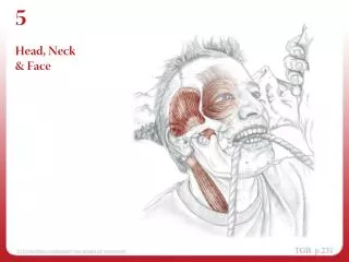
Anatomy: Skull • Consists of 8 cranial bones and 14 facial bones • Brain (encephalon) is housed in the cranium • Afforded considerable protection via an ingenious system of bony and soft-tissue structures
Anatomy: Skull • Bones of the cranium • Are held together by specialized articulation known as suture joints • Do not complete their ossification process until human beings are between 20 and 30 years old (Gray, 1985) • Soft-tissue structure serve as protective function include the five layers of tissues • Skin – layer of dense connective tissue • Galeaaponeurotica –broad, flat tendon • Loose connective tissue • Periosteum of the cranial bone
Anatomy: Meninges • Below the cranial bones a group of soft tissue is found called the cerebral meninges • 3 distinct layers of tissue between the underside of the cranium and the surface of the brain • Dura mater • Outermost • Tough fibrous connective tissue that function as a periosteum to inside surfaces of cranial bones • Protective membrane • Highly vascular, has both arteries and veins transport blood to and from the cranial bones
Anatomy: Meninges • Arachnoid • Middle layer • Less strength and contains no blood supply • Pia Mater • Inner most layer • Physically attached to the brain tissue and serves to provide a framework for an extensive vasculature that supplies the brain • Very thin, delicate membrane • More susceptible to trauma than the dura mater • Subarachnoid space • Containing cerebrospinal fluid (CSF) • Cushion the brain and spinal cord from external forces • Like collision and contact sports
Anatomy: Central Nervous System • The brain (cencephalon) along with the spinal cord compose the central nervous system (CNS) • Brain and spinal cord are protected by the meninges and bony structure of the cranium and vertebrae • Consists of gray and white matter that represent two distinct types of neural tissues • Weights 3 to 3.5 lbs contains 100 billion neurons
Anatomy: Central Nervous System • 3 basic parts • Cerebrum • Largest of the three • Involved in complex functions like cognition, reasoning, and intellectual functioning • Cerebellum • Lower posterior portion • Performs functions related to complex motor skills • Brain stem • Base of the brain • Connects the brain to the spinal cord
Anatomy: Face • Composed of an outer layer of skin • Facial bones • Maxilla (upper jaw) • Right and left palatine • Right and left zygomatic • Right and left inferior nasal concha • Vomer • Mandible (lower jaw) • hyoid
Anatomy: Face • Several areas around the face are especially prominent thus prone to injury • Orbits for the eyes (contusions) • Nasal bones (fractures) • Lower jaw (mandible) • Excessive external forces
Anatomy: Cervical Spine • 7 cervical vertebral • The first cervical 9C-1) vertebra (atlas) articulates directly with the occipital bone to form the right and left atlantoccipital joints • The skull and C-1 articulate as a unit with the second cervical (C-2) vertebra (axis) to form the atlantoaxial joint • Allows for rotation of the head on the neck • The remaining 5 cervical vertebrae become larger as they approach the thoracic spine
Head Injuries in Sports • Relatively minor trauma to the head can result in severe, sometimes life-threatening injury • Inability of brain tissue to repair itself, any loss of tissue results in some level of permanent disability • If severe enough can result in death • With appropriate education, coaches can learn to recognize head injuries and render effective first aid when necessary
Head Injuries in Sports • Surveys have provided additional insight into which sports appear to carry a higher risk • Guskiewicz and colleagues (2000) conducted a 3-year study of head/brain injuries in the US among high school and collegiate football players • Overall they estimated that approximately 300,000 traumatic head or brain injuries happened annually • Players sustaining a concussion had a threefold increased risk of sustaining an additional concussion
Head Injuries in Sports • When examining high school and collegiate female activities, cheerleading leads the list of activities resulting in directly related catastrophic injuries • Attributed to the escalated degree of difficulty in cheerleading routines as it has become a competitive sport • 26,786 hospital emergency room visits related to cheerleading injuries occurred in 2007 • 783 concussions • 308 contusions • 69 lacerations • 1122 internal injuries 9Mueller & Cantu, 2010)
Head Injuries in Sport • All head injuries can be placed into three general categories: • mild head injury or concussion • intracranial hemorrhage • skull fracture
Mechanism of Injury • Involve either direct or indirect mechanisms • Direct • Blow to the head resulting in brain injury at site of impact • Coup type injury – site of maximal injury is usually at the point of impact • Ex) accelerated force is generated when an opponent or the ball hits an athletes head • Countercoup type injury – the site of maximal injury is opposite the point of impact • Ex) deceleration forces are generated when an athlete’s head strikes the ground • Indirect • Damaging forces traveling from other areas of the body, such as blows to the face or jaw • Rapid and violent movement of the cervical spine (whiplash) • Treat every head injury as if there is a neck injury, and every neck injury as if there is also a head injury
Coup vs. Counter Coup Coup = site of maximal injury is at the site of impact (opponent or the ball hits an athletes head Counter Coup – site of maximal injury is opposite the point of impact (the brain rebounds against the skull )
Concussion (Mild Head Injury) • Defined by Jordan (1989) as a clinical syndrome characterized by immediate and transient impairment of neurologic function secondary to mechanical forces • Clinical manifestations • Headache • Dizziness • Confusion • Unconsciousness • Inability to quickly answer questions about orientation • Irritability • Poor concentration • Pupils react to light • Poor ability to track with eyes • Poor depth perception • Ringing in the ears • Vomiting • Nausea • Actions uncharacteristic of the individual
Concussion (Mild Head Injury) • Recent evidence suggest in some concussion there is a level of structural damage • Brain cells are not destroyed remain extremely vulnerable to subsequent trauma • Result in minor changes in blood flow • Intracranial pressure • Anoxia (Cantu, 2001) • The majority base the level of severity on duration of unconsciousness as well as the presence or absence of post-traumatic amnesia (PTA) • Extremely difficult to gauge the length of time a person is unconscious
Cantu Evidence-Based Grading System for Concussion 3 Grades: Grade 1 (mild) Grade 2 (moderate) Grade 3 (severe)
Concussions (Mild Head Trauma) • Grade 1 are the most difficult to identify • Grade 3 are distinct because they involve either a loss of consciousness lasting more than 1 minute or PTA of greater than 24 hours • Majority of sports-related concussions involve periods of unconsciousness lasting 1 minute or less
Concussions (Mild Head Trauma) • Two types of PTA: • Anterograde Amnesia • Involves an inability to recall events that have transpired since the time of the injury • Retrograde Amnesia • Present when the athlete is unable to recall events that occurred just prior to the injury • It is generally thought that retrograde amnesia is indicative of more severe forms of head injury
Concussions (Mild Head Trauma) • Level of consciousness • First determining if the athlete is alert and will respond to simple questions • Keep questions simple to evaluate the athlete’s perspective of time and place • Ex) game score, name of opponent, reciting four words or numbers immediately and 2 minutes later to detect the presence of anterograde amnesia (Cantu, 2001) • Remember consciousness does not guarantee the absence of a potentially serious head injury
Second Impact Syndrome (SIS) • “Occurs when an athlete who has sustained an initial head injury, most often a concussion, then sustains a second head injury before symptoms associated with the first have fully cleared” (Cantu & Voy, 1995) • Rapid development of catastrophic swelling of the brain • Puts pressure directly against the brain stem
Intracranial Injury • Is a potentially life-threatening situation • Direct blows, rapid deceleration, and even rapid rotational motions of the head • Result from blunt trauma to the head • Characterized by disruption of blood vessels either veins or arteries result in the development of a hematoma or swelling in the confines of the cranium
Intracranial Injury • Epidural hematoma • Bleeding between the dura and the cranial bones • Involves arterial bleeding • Signs and symptoms of injury will develop rather quickly • Subdural hematoma • Bleeding below the dura mater • Involve rapid arterial bleeding • Symptoms develop in minutes • Or pooling and clotting develop over many hours • Intracerebral hematoma • Bleeding within the brain tissues • Cerebral contusion • Bruising of the brain tissue
Cranial injury • Involve the bones of the skull • May be come bleeding and soft-tissue damage • More severe forms of cranial injuries involve depressed skull fractures • more serious because bone fragments have been pushed into the cranial region • More likely to produce serious, perhaps life-threatening, neurologic damage
Initial Treatment of a Suspected Head Injury: Guidelines • Divided into procedures while the athlete is at the site of injury • If any signs and/or symptoms of head and neck injury are present when evaluating the athlete at the site of initial injury, he/she should not be moved until emergency medical (EMS) personnel have arrived on site
Initial Check • First step • Incorporates basic first aid procedures • Determine if athlete is either respiratory or cardiac arrest • Accomplished by executing the initial check • Obstructed airway or cardiac arrest must be attended to before continuing • First few seconds provide important information • Note body position, movement or lack thereof, unusual limb positions and (if present) the position of helmet, face mask, and mouth guard
Initial Check • Unconscious • Awake him/her by placing hands on the shoulders, chest, or upper back and speaking loudly directly toward the athlete's head • Make a mental note of time • Conscious • Airway is probability open • Great importance that the coaching staff be trained and well rehearsed in dealing with such situations • Immobilization of the head and neck should also take place at this time
Initial Check Person stationed at the athlete's head to stabilize it with both hands Normally the ATC
Initial check • In case of a helmeted football player it is not necessary to remove the helmet to determine breathing • Usually be detected by placing an ear near the athlete’s face and listening for sounds of respiration • Detect sounds indicating airway obstruction such as gagging, wheezing, or choking
Circulation Assessment • Responsive athlete who is breathing will have signs of circulation • Breathing • Coughing • Movement • Determine if signs of circulation are present • If no signs are present, can begin cardiopulmonary resuscitation (CPR) and activate the EMS provider
Circulation Assessment • Proper guidelines for spine boarding • 1st the captain of the team stabilizes the head and neck in the exact position in which they were found • Place the arms next to the body and legs straight • If lying face down roll the athlete supine • Four or five people are required to “log roll” • Position the arms in the cross arm technique • 2nd place spine board as close as possible beside individual • Each person is responsible for one body segment: one at the shoulder, one at the hip, one at the knees, and if needed one at the feet • On command roll the individual on the board in a single motion • 3rd once on board the captain continues to stabilize the neck and head • Coach’s primary responsibility is to keep the athlete alive and ensure help is summoned • No need to remove the athlete
Physical Exam • Once initial check is completed and the athletes vital signs have been ascertained, proceed to the physical exam • C - conscious or unconscious • E – extremity strength (if conscious) • M – mental function (if conscious) • E – eye signs and movements • P – pain specific to neck • S – spasm of neck musculature • DON’T REMOVE THE HELMET OF A FOOTBALL PLATER • DON’T MOVE THE ATLETE • DON’T RUSH THROUGH THE PHYSICAL EXAM
Physical Exam • If seem to be conscious, attempt to communicate by asking simple questions • Name of opposing team, what day it is, location of the contest • Not attempt to revive an unconscious athlete by using a commercially made inhalant • Ammonia capsules • Athlete may jerk head result in aggravation of existing neck injury
Physical Exam • If conscious can conduct a series of quick, simple test to determine if any significant neurologic damaged has occurred • Grip strength test • Place hands on tops of athletes feet and ask player to dorsiflex • Compare bilateral strength • Check dermatomes or myotomes
Sideline Assessment • A more detailed assessment of his/her condition • Determine if any S&S of head injury have developed since time of initial injury • Single most important indicator of severity of brain injury is level of consciousness (Jordan, 1989) • Use Standardized Assessment of Concussion (SAC) • Administered at the site of injury • Has sustained a concussion but – loss of consciousness (LOC)
SAC • Assess orientation, immediate memory, concentration and delayed recall (McCrea et al., 1998) • Asking a series of questions • Recall five words • Motor skills include push-ups, knee bends, sit-ups, and jumping jacks • Use SAC over the course of the athletes recovery in conjunction with a physicians care
Cervical Spine Injuries • Neck (cervical) injuries occur in almost any sport • Extent and severity of neurologic damage depends on the magnitude of the mechanism of injury, resulting movement of the neck, and the extent of tissue damage • More serious • Displacement of an intact vertebra occurs • Fragments of a vertebral fx are displaced • Intervetebral disk ruptures • Placing pressure directly on the spinal cord or nerve roots