Schwann Cells
170 likes | 1.23k Views
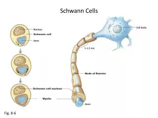
Schwann Cells. Cell body . Nucleus. Schwann cell. Axon. 1 – 1.5 mm . Node of Ranvier. Schwann cell nucleus. Myelin. Axon. Fig. 8-6. General structure of neurons. Input signal. Dendrites. Integration. Cell body. Nucleus. Axon hillock. Axon (initial segment). Myelin sheath.

Schwann Cells
E N D
Presentation Transcript
Schwann Cells Cell body Nucleus Schwann cell Axon 1–1.5 mm Node of Ranvier Schwann cell nucleus Myelin Axon Fig. 8-6
General structure of neurons Input signal Dendrites Integration Cellbody Nucleus Axon hillock Axon (initialsegment) Myelinsheath Presynapticaxon terminal Outputsignal Synapticcleft Synapse Postsynapticdendrite Postsynapticneuron Fig. 8-2
Chemical synapses – General structure Axon ofpresynapticneuron Mitochondrion Axon terminal Postsynaptic neuron Synapticvesicles Synapticcleft Neurotransmitter Receptors Fig. 8-20
Axon terminalsof presynapticneurons Dendrite ofpostsynapticneuron Glial cellprocesses Axon Fig. 8-26
Stimulus Stimulus Stimulus Enclosed nerveending Free nerve endings Specialized receptorcell (hair cell) Layers of connectivetissue Synaptic vesicles Synapse Unmyelinatedaxon Myelinated axon Myelinated axon Cell body Cell body Cell body ofsensory neuron Fig. 10-1