Download
1 / 8
80 likes | 242 Views
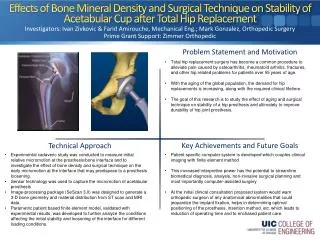
Effects of Bone Mineral Density and Surgical Technique on Stability of Acetabular Cup after Total Hip Replacement. Investigators: Ivan Zivkovic & Farid Amirouche , Mechanical Eng.; Mark Gonzalez, Orthopedic Surgery Prime Grant Support: Zimmer Orthopedic.

E N D
Effects of Bone Mineral Density and Surgical Technique on Stability of Acetabular Cup after Total Hip Replacement Investigators: Ivan Zivkovic & FaridAmirouche, Mechanical Eng.; Mark Gonzalez, Orthopedic Surgery Prime Grant Support: Zimmer Orthopedic • Total hip replacement surgery has become a common procedure to alleviate pain caused by osteoarthritis, rheumatoid arthritis, fractures, and other hip related problems for patients over 55 years of age. • With the aging of the global population, the demand for hip replacements is increasing, along with the required clinical lifetime. • The goal of this research is to study the effect of aging and surgical technique on stability of a hip prosthesis and ultimately to improve durability of hip joint prosthesis. • Experimental cadaveric study was conducted to measure initial relative micromotion at the prosthesis/bone interface and to investigate the effect of bone density and surgical technique on the early micromotion at the interface that may predispose to a prosthesis loosening. • Sensor technology was used to capture the micromotion of acetabular prosthesis • Image-processing package (SeScan 3.0) was designed to generate a 3-D bone geometry and material distribution from ST scan and MRI data. • Parametric patient based finite element model, validated with experimental results, was developed to further analyze the conditions affecting the initial stability and loosening of the interface for different loading conditions. • Patient specific computer system is developed which couples clinical imaging with finite element method • This increased interpretive power has the potential to streamline biomedical diagnosis, analysis, non-invasive surgical planning and most importantly computer-assisted surgery • At the initial clinical consultation proposed system would warn orthopedic surgeon of any anatomical abnormalities that could jeopardize the implant fixation, helps in determining optimal positioning of the prosthesis, insertion method, etc. which leads to reduction of operating time and to enchased patient care.
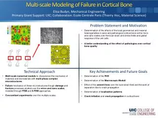
Multi-scale Modeling of Failure in Cortical Bone Investigator: Elisa Budyn, Mechanical Engineering Grant Support: UIC; Collaboration: EcoleCentrale Paris (Thierry Hoc, Material Science) • Determination of the effects of the local geometrical and material heterogeneities in sane and pathological cortical bone at the micro and nano scales over the local strain and stress fields and global response of the unit cells. • A better understanding of the effect of pathologies over cortical bone quality • Determination of the RVE • Determination of the Macroscopic Moduli • Effect of the cement lines over the local strain field and the work of separation due to crack propagation • Determination of localization patterns • Crack initiation and crack propagation in cortical bone • Multi-scale numerical models to characterize the mechanics of materials and biomaterials with multi-phase complex microstructures. • Failure mechanics of these microstructures though damage and fracture processes studied over the micro and nano scales, modeled through FEM and X-FEM approaches. • Concomitant experiments over the multiple scales.
Orienting Human Stem Cells (hMSCs) by Means of Electrospun Polymer Nanofibers Investigators: M. Cho, Bioengineering; A. Yarin, C. M. Megaridis, Mechanical and Industrial Engineering; E. Zussman, Technion-Israel Oriented Random • Cell orientation and adhesion control the functionality of natural and engineered tissues • Electrospinning is a low-cost technique which can produce polymer nanofibers aligned along a specific direction • Polymer nanofibers can be used to mimic the native extracellular matrix (ECM) features • Electrospun polymer nanofiber scaffolds are used to manipulate cell orientation and adhesion Cells: Green, Nanofibers: Red • Random and oriented polycaprolactone (PCL) nanofibrous scaffolds produced using electrospinning • hMSCs were cultured and seeded on two scaffold types (random, oriented) • Orientations of hMSCs and nanofibers on random and oriented nanofibrous scaffold samples were measured via laser scanning confocal microscopy at different time points during an 18-day culture period • hMSC viability tests were performed to verify compatibility of the cells with the PCL • hMSCs adhered and oriented along PCL nanofibers • During long-term culture, hMSCs demonstrated no preferred orientation on random nanofibrous scaffolds; cells consistently aligned on oriented scaffolds • Oriented PCL nanofibrous scaffolds could be used to mimic the cell and ECM organization in the native tissue, such as muscle, tendon, and the superficial zone of articular cartilage • The fiber scaffold/hSMC approach holds promise for a variety of tissue engineering applications
Artery Fluid flow Noise generation Approximate location of constriction Multimode Sonic & Ultrasonic Diagnostic Imaging Investigators: Thomas J. Royston & Francis Loth, Mechanical & Industrial Engineering Prime Grant Support: NIH • Ultrasonic (US) imaging provides detailed geometry of blood vessels • Geometric changes may indicate disease or injury • Sonic imaging provides unique functional information • Sounds associated with disease are sonic, not US • Merge US and Sonics to harness strengths of each • Initial application: peripheral vascular pathologies – vessel constrictions (plaque and intimal hyperplasia) Blood vessel with constriction in soft tissue phantom Grayscale of geometry from US imaging Color overlay of acoustic field generated by turbulence downstream of the constriction • Sonic wave propagation in biological tissue is more complex than US • . • Requires new acoustic modeling developments • Inverse modeling to extract acoustic image from array • Novel acoustic sensor development • Merging multiple imaging modalities on same platform • Prototype US/Sonic system has been developed: • conventional US system retrofitted with • electromagnetic position device for true 3D imaging • acoustic sensor array pad that is transparent to US so US imaging can be conducted with the pad in place • Calibration of system on phantom models in progress • Turbulence imaged downstream of vessel constriction • Future plans: Human subject studies, improved prototype, better sensor array, improved imaging software Prototype 15 sensor sonic array pad on arm
The Audible Human Project Investigator: Thomas J. Royston, Mechanical & Industrial Engineering, Bioengineering Primary Grant Support: NIH • Develop and experimentally validate a subject-specific computer model of sound generation, transmission and measurement in the pulmonary system and chest. • Motivation: Complement to National Library of Medicine “Visible Human Project.” Research and education/ training tool. Integration into Haptic Virtual Reality environment in the future (e.g. ImmersiveTouch™). Mechanical phantom model for code validation: foam with airways (lungs) surrounded by silicon with embedded garalite ribs (chest wall). Wire mesh geometry of chest surface, lungs and main airways based on Visible Human Male. • Patient-specific acoustic model based on coupling an analytical airway model with a lung tissue boundary element model and finite element model of the ribcage and chest surface • Validated via experimental studies on phantom models and human subjects • Code validation via experimental phantom studies in progress • Development of computational model based on Visible Human Male in progress • Future plans: Experimental validation on human subjects • Future plans: Extend to cardiovascular, musculoskeletal and gastroinstestinal systems • flexible sonic sensor array pad
Biomimetic MEMS Technology for a Novel Retinal Prosthesis PI: LaxmanSaggere, Mechanical and Industrial EngineeringCollaborator: David Schneeweis, BioEngineering Prime Grant Support: National Science Foundation • Motivation: Photoreceptor degeneration in diseases such as ARMD and RP is the leading cause of blindness in the world. No cures or therapies are available for these diseases, but a retinal-based prosthesis offers a promising treatment option. Most current retinal prostheses rely on the concept of electrical stimulation of neurons, which is conceptually simple, but faced with many challenges • Objective: To develop a biomimetic technology enabling a fundamentally different and technically superior approach to a retinal prosthesis. This approach, in principle, mimics a natural photoreceptor’s function of transducing visual stimuli into chemical signals that stimulate the surviving retinal neurons. • Approach: A microdispenser unit integrated with a miniaturized solar cell and a thin-film piezo actuator on one side and several micron-scale ports on the other side contains liquid chemical (neurotransmitter). An array of such microdispenser units constitutes the core of a prosthesis. • Principle of Operation: Light falling on the retina irradiates the solar cell, which generates voltage across the piezo actuator. The actuator pressurizes the liquid and dispenses it through the micro ports. The liquid diffuses through micro-capillaries in a soft encapsulation and stimulates retinal cells. • Technologies: MEMS, microfluidics, thin-film piezoelectric actuators, solid-sate solar cells, chemical cellular signaling. • Challenges: i)Low intensity light at the retina; ii) Integration of array components and microfluidics; iii) Chemical dispensing rate, mechanism, long-term operation; iv) Biocompatible packaging. • Key Achievements:i) Completed preliminary system design and established the concept feasibility; ii) Established a technique to chemically stimulate neuronal cells and record the cellular response; iii) Fabricated and characterized the light powered actuator; iv) Established techniques to quantify nanoliter flow • Future Goals:i) To fabricate and test an in-vitro proof of the concept device; ii) To lead the technology developed towards clinical relevancy through interdisciplinary collaborations with neuroscientists and retina specialists.
Universal Design of Exercise Equipment for People with Disabilities Investigators: Michael J. Scott, Mechanical & Industrial Engineering Primary Grant Support: U.S. Dept. of Education (OSERS/NIDDR) : RERC RecTech • Lack of access to exercise is a major health risk for people with disabilities • Wheelchair users are particularly challenged to find appropriate cardiovascular exercise; the common arm ergometer is a risk of shoulder overuse injury • Major equipment manufacturers and gyms have limited interest in what they perceive as a niche market • Regulation and standards driving the push for more universal equipment • Consider physiological requirements and usability needs first • Mechanism design to permit universally designed machines that serve the exercising population both with and without disabilities • Partnership with Life Fitness • Collaboration with investigators at SUNY Buffalo developing instruments to measure universality of products • Collaboration with standards developers in the United States (Beneficial Designs) and Great Britain (Inclusive Fitness Initiative) • Categorized and identified best candidate exercise motions for wheelchair users with different levels of function to achieve cardiovascular benefit without risk of overuse injury • First prototype of dual-use adapted Life Cycle 9500HR currently being tested on human subjects by colleagues Thayne Munce of Movement Sciences and Karen Troy of Kinesiology and Nutrition • Rehabilitation Engineering Research Center (RERC) RecTech funding renewed through 2012 • Future developments: adaptation of strength equipment for cardiovascular use
Stimuli-Responsive Polymer Nanofibers Y. Zhang, Prof. A. L. Yarin Mechanical and Indutrial Engineering, UIC • Water insoluble novel NIPAM-based copolymers • Swelling/shrinkage in response to temperature variation • Swelling/shrinkage in response to pH variation • Controlled drug release • Triggering at pH of 6.5 characteristic of cancer tumors • Water insoluble novel NIPAM-based, thermo- and pH-responsive copolymers were synthesized • These copolymers can distinguish between cancer tumors (pH 6.5) and normal tissues (pH 7.4) and release an anti-cancer drug in a highly localized manner, thus eliminating severe side effects • Future experiments should involve real anti-cancer drugs • Drug delivery with nanobots: carbon nanotubes containing anti-cancer drugs and capped with these stimuli-responsive copolymers • Co-polymerization of thermo-responsive NIPAM-PMMA copolymers • Co-polymerization of pH-responsive NIPAM-PMMA-AA copolymers • Electrospinning of nanofiber mats loaded with a model compound-fluorescent dye • Thermo- and pH-activated periodic dye release • To appear in: Y. Zhang, A. L. Yarin, J. Materials Chemistry (2010)