Download
1 / 7
280 likes | 3.98k Views
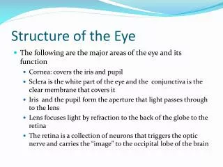
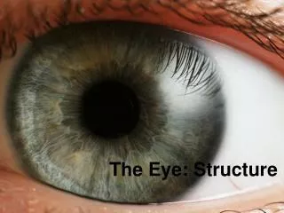
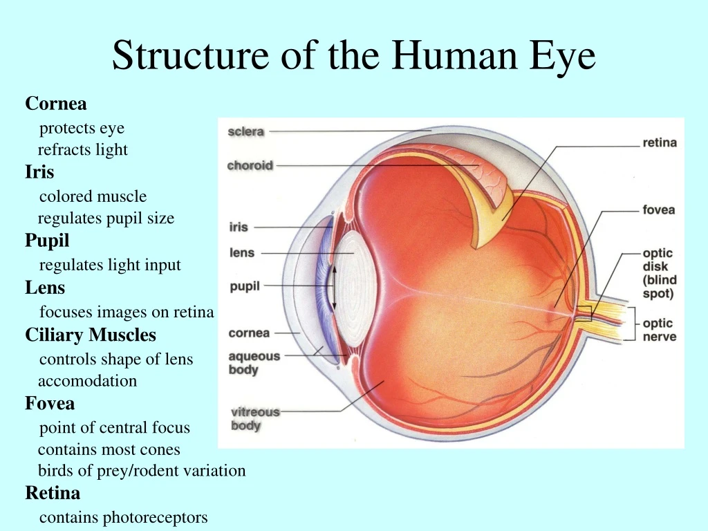
Structure of the Human Eye. Cornea protects eye refracts light Iris colored muscle regulates pupil size Pupil regulates light input Lens focuses images on retina Ciliary Muscles controls shape of lens accomodation Fovea point of central focus contains most cones

E N D
Structure of the Human Eye Cornea protects eye refracts light Iris colored muscle regulates pupil size Pupil regulates light input Lens focuses images on retina Ciliary Muscles controls shape of lens accomodation Fovea point of central focus contains most cones birds of prey/rodent variation Retina contains photoreceptors
The Retina (make up the optic nerve) • Rods • 100-120 million • sensitive to dim light • black/white discrimination • large numbers on the periphery • Cones • 4-6 million • used for color vision • located near the fovea • red, green, and blue cones
Visual Pathway Light to rods/cones to bipolar cells to ganglion cells to LGN cells to Visual Cortex
Bipolar cell Active Not Active Photoreceptor Action • In the Dark: • rods are depolarized • rods release glutamate • glutamate is inhibitory • bipolar cells are inhibited • In the Light: • rods are hyperpolarized • no glutamate is released • bipolar cells are not • inhibited (disinhibition) • bipolar cells undergo • spontaneous activity Glutamate (-) Rod cell LIGHT DARK
Rhodopsin Photopigment Rhodopsin: made up of retinal and opsin spans the disc membrane acts as a G-protein
Light Transduction LIGHT DARK • trans-retinal transformed to cis-retinal • cis-retinal and opsin form rhodopsin • rhodopsin activates guanylate cyclase (GC) • GC increases the synthesis of cGMP • cGMP opens Na+ channels • rod cell depolarizes • increases the release of glutamate • (darkness adjustment–waiting for rhodosin) • cis-retinal transformed to trans-retinal • trans-retinal and opsin dissociate • now active opsin activates transducin • transducin activates PDE • PDE breaks down cGMP to 5’-GMP • 5’GMP closes Na+ channels • rod cell hyperpolarizes • reduces the release of glutamate
Rhodopsin Cascade Rhodopsin molecule LIGHT Rod cell disc Inside Rod cell Outside 1 photon of light can block the entry of 1,000,000 Na+ ions