Download
1 / 30
340 likes | 697 Views
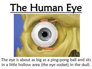
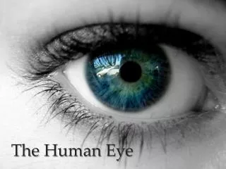
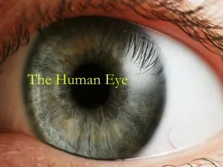
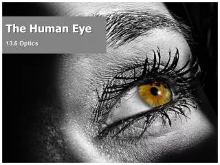
The Human Eye. 13.6 Optics. 13.6. THE HUMAN EYE: How Images Are Formed. The human eye gathers light from objects. In a healthy eye, a smaller, inverted, real image of an object is created on the retina at the back of the eye.

E N D
The Human Eye 13.6 Optics
13.6 THE HUMAN EYE:How Images Are Formed • The human eye gathers light from objects. • In a healthy eye, a smaller, inverted, real image of an object is created on the retina at the back of the eye. • Electrical impulses from the eye travel through the optic nerve to the brain. The brain • Takes inverted image from the retina and flips it so that the image we “see” appears upright
In a healthy eye, a smaller, inverted, real image of an object is created on the retina at the back of the eye.
Eye muscles called ciliary muscles slightly change the shape of the eye lens. • The process of accommodation makes it possible to create a sharply focused image on the retina if an object is distant or nearby.
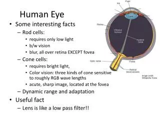
Parts of the Human Eye Iris • Coloured part of eye • Opens and closes around a central hole to controls the amount of light that gets in Pupil • The hole in the iris
Parts of the Human Eye Cornea & Lens combination • Cornea – transparent bulge over the lens • Together, acts like a converging lens • Produces a smaller, real, inverted image on the retina
Parts of the Human Eye Retina • Light sensitive cells in the retina convert light signals into electrical signal that is transmitted to the brain through the optic nerve Optic Nerve • Creates blind spot at the back of each eye (but is compensated for by the other eye)
Perceiving and Seeing • The retina is an extension of the brain and consists of several complex layers of nerve cells. • Light from different directions are received by different parts of the retina because the retina is not perfectly consistent in terms of the sharpness of the image we perceive. • The fovea is the are in the center of the field of view which produces the clearest image. • There is also something called the blind spot in the eye and this is where the nerves carrying all the information leave the eye in a narrow bundle. TRY IT!!
Perceiving vs. seeing in the brain • The physical task of forming an image is only one part of the human perception of light. • In order to see, the light entering your eyes must be absorbed by photoreceptors, light sensitive cells found in the retina that are shaped as rods or cones. • Rod cells allow for the detection of shapes and movement in low light and only shades of grey are detected by these cells. • Cone cells are the photoreceptors that detect three primary colours: red, green, or blue.
Accommodation • Our ciliary (eye) muscles help the eye focus on distant and nearby objects by slightly changing the shape of the eye lens, thereby changing the focal length to allowing the eye to focus the image on the retina • The process of accommodation makes it possible to create a sharply focused image on the retina if an object is distant or nearby.
Focusing Problems Hyperopia (far-sightedness) • Can see far / can not see nearby objects • Cause: • distance between lens and retina too small, or • cornea-lens combination too weak
Hyperopia (far-sightedness) • Result:light from all nearby objects focuses behind the retina
Hyperopia (far-sightedness) • Solution: • Far-sighted eyes need help refracting light • a corrective converging lens (with a positive meniscus)
Focusing Problems Presbyopia • A form of far-sightedness caused by a loss accommodation as a person ages • Eye lens just loses its elasticity
Focusing Problems Myopia (near-sightedness) • Can not see far / can see nearby objects • Cause: • Distance between lens and retina is too large, or • Cornea-lens combination converges light too strongly
Focusing Problems • Result: light from distant objects is brought to focus in front of the retina
Focusing Problems • Solution: a corrective diverging lens (with a negative meniscus)
Focusing Problems Astigmatism • abnormal curvature of the cornea (oval shape) can cause two focal points to fall in two different locations making objects up close and at a distance appear blurry
Focusing Problems Glaucoma • the eye’s drainage system becomes clogged so the intraocular fluid cannot drain. As the fluid builds up, it causes pressure to build within the eye. High pressure damages the sensitive optic nerve and results in vision loss.
Focusing Problems Cataracts • A clouding of the eye’s natural lens • Blocks light from reaching the retina and interferes with vision • Cataract Surgery
Eye Floaters (Savanna!) • Eye floaters are small moving spots that appear in your field of vision. They may be especially noticeable when you look at something bright, such as white paper or a blue sky. • Most of the time people learn to live with eye floaters and ignore them. And they often improve over months to years. Only rarely do benign eye floaters become bothersome enough to consider treatment. • But sometimes eye floaters are a sign of a more serious condition. You should seek immediate medical attention if you notice a sudden increase in the number of eye floaters.
Eye floaters move as the eyes move. They generally appear to dart away when you try to focus on them. • Eye floaters can appear in many different shapes, such as: • Black or gray dots • Squiggly lines • Threadlike strands, which can be knobby and semi-transparent • Cobwebs • Ring shaped
Causes of Eye Floaters • Most eye floaters are caused by small flecks of a protein called collagen. • The back compartment of the eye is filled with a gel-like substance called vitreous humor. • As you age, the vitreous and its millions of fine collagen fibers shrink and become shred-like. Shreds can accumulate in the vitreous. This can cause a change in the amount of light that hits the These changes can happen at any age. They most often occur between ages 50 and 75, especially in people who are very nearsighted or have had cataract surgery.
Eye Colour What Eye Colour will you Children Have? • http://genetics.thetech.org/online-exhibits/what-color-eyes-will-your-children-have Eye Colour • http://www.eyedoctorguide.com/eye_general/eye_color_genetics.html