Download
1 / 25
330 likes | 832 Views
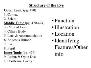
Outer Tunic (pg. 470) 1. Cornea 2. Sclera Middle Tunic (pg. 470-474) 3. Choroid Coat 4. Ciliary Body 5. Lens & Accommodation 6. Aqueous Humor 7. Iris 8. Pupil Inner Tunic (pg. 474) 9. Retina & Optic Disc 10. Posterior Cavity. Structure of the Eye. Function Illustration

E N D
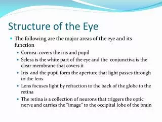
Outer Tunic (pg. 470) 1. Cornea 2. Sclera Middle Tunic (pg. 470-474) 3. Choroid Coat 4. Ciliary Body 5. Lens & Accommodation 6. Aqueous Humor 7. Iris 8. Pupil Inner Tunic (pg. 474) 9. Retina & Optic Disc 10. Posterior Cavity Structure of the Eye • Function • Illustration • Location • Identifying Features/Other info
12.6: Structure of the Eye 2.1 Atoms, Ions, and Molecules Sponge: Set up Cornell Notes on pg. 69 Topic: 12.6: Structure of the Eye Essential Questions: • How does the shape of the lens change during accommodation?
Pg. 68 Human Body: Pushing the Limits: Sight • Take at least 15 bullet points
Directions: • You will need 12 colors for your table • As we go through each part of the notes, please color-code and label the “diagram of the eye” AND your Brace Map notes • Also, add any missing info.
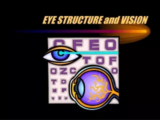
Structure of the Eye Pg. 69 • Hollow • Spherical (2.5 cm diameter) • Wall has 3 layers • outer fibrous tunic • middle vascular tunic • inner nervous tunic
Outer Fibrous Tunic • Cornea • Anterior 1/6 of outer eye • “Window of the eye” • Focuses incoming light rays • Transparent • No blood vessels • Well supplied with nerves • Many pain receptors
Outer Tunic • Sclera • Posterior 5/6 of outer eye • White portion of eye • Protects the eye • Attachment for extrinsic muscles • Optic nerve pierces the sclera in the back
Middle Tunic • Choroid coat • Many blood vessels • provides blood supply • Many melanocytes • pigments absorb extra light • Keeps inside of eye dark
Middle Tunic • Ciliary body • Anterior portion of middle tunic • Holds lens in position • Moves lens • Secretes aqueous humor into the posterior chamber
Middle Tunic • Lens • Lies behind iris and pupil • Elastic • Under constant tension • Puts near/far objects into focus • As we age, lens becomes larger and less elastic which leads to vision impairment Accommodation: Suspensory ligamentsCiliary ligaments Close viewing relaxed contracted Far Viewing contracted relaxed
Figure 12.29 Accommodation • changing of lens shape to view objects
Aqueous humor (a-quee-us): fluid that circulates through the pupil and into the anterior chamber of the eye • Provides nutrients • Maintains the shape of the front of eye • Removes waste • If drainage is blocked—leads to glaucoma which can result in blindness
Middle Tunic • Iris • Colored portion of eye • Lies between cornea and lens • Smooth muscles control the size of the pupils • Pupils • Controls light intensity • Constricted: less light in • Dilated: more light in
Iris Pupil
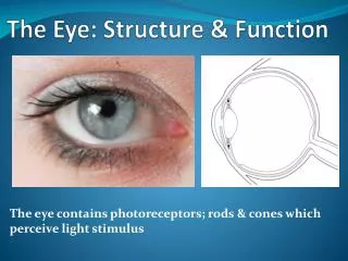
Inner Tunic • Retina • Contains visual receptors (photoreceptors) rods/cones • Continuous with optic nerve • macula lutea (mac-ulalu-tay-a)–spot in retina; absorbs extra light • fovea (fo-vea) centralis– center of macula lutea; region of retina that produces the sharpest vision; has largest concentration of cone cells • optic disc – Blind spot; contains no visual receptors • Nerve fibers leave here and become part of the optic nerve
Retina Optic Disc Optic Nerve
Inner Tunic • Posterior Cavity • largest compartment in eye • contains vitreous (vi-tree-us) humor – jelly-like substance that holds retina flat against choroid coat maintaining pressure • Helps transmit light to retina
Video Notes: Split pg. 69 into four sections • Outer fibrous tunic • Middle vascular tunic • Inner nervous (sensory) tunic • The Humors and the Lens • 3-5 Bullets Each