Download
1 / 19
190 likes | 475 Views
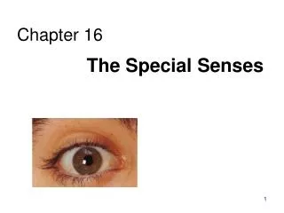
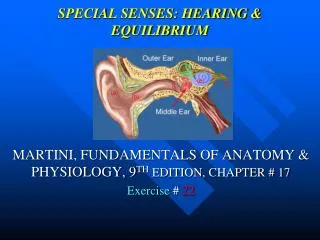
SPECIAL SENSES: HEARING & EQUILIBRIUM. MARTINI, FUNDAMENTALS OF ANATOMY & PHYSIOLOGY, 9 TH EDITION, CHAPTER # 17 Exercise # 22. NOTE:. THIS IS A STUDY GUIDE , NOT AN ALL INCLUSIVE REVIEW.

E N D
SPECIAL SENSES: HEARING & EQUILIBRIUM MARTINI, FUNDAMENTALS OF ANATOMY & PHYSIOLOGY, 9TH EDITION, CHAPTER # 17 Exercise # 22
NOTE: • THIS IS A STUDY GUIDE, NOT AN ALL INCLUSIVE REVIEW. • THERE MIGHT BE THINGS NOT COVERED BY THIS STUDY GUIDE THAT MIGHT BEASKED IN YOUR PRACTICUMS / QUIZZES. • STUDENTS ARE RESPONSIBLE FOR READING THEIR TEXBOOK (S) AND FOR ALL THE MATERIAL COVERED DURING THE LABORATORY PERIOD, AS PER THE COURSE SYLLABUS
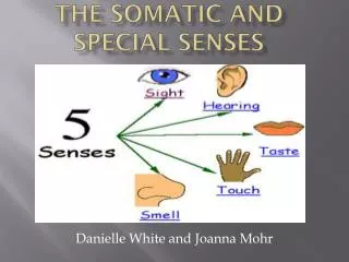
OBJECTIVES • Identifying the structures of the ear and describe their functions.
EXTERNAL EAR MIDDLE EAR INNER, OR INTERNAL EAR
EXTERNAL EAR • Pinna or auricle- to capture the sound waves • External auditory canal-to focus & direct the sound waves • Ceruminous glands (lining the external auditory canal) • f- to produce waxy material • To avoid foreign matters to go inside • Tympanic membrane- to transmit the sound waves
MIDDLE EAR • Ossicles(3 little bones)- malleus, incus & stapes • F- to transmit the sound waves • Pharyngotympanic or auditory or eustachian tube • F- to equalize the pressure inside & outside the • tympanic membrane
INNER OR INTERNAL EAR • The bony labyrinth -it has 3 parts- vestibule, • semicircular canal & cochlea • It surrounds the membranouslabyrinth • It contains the perilymph • Perilymph (pink in model)-to transmit sound waves • Membranous labyrinth (grey in model)- it Contains the endolymph • Endolymph- to transmit sound waves
Vestibule- balance & equilibrium • It contains the utricle & sacule • Utricle & sacule- contain themacula • F- sensations of gravity & linear acceleration • Semicircular canals- sense of dynamic balance • Ampulla- swollen area at the end of the • Semicircular canal that contains the Crista • Crista- contains sensory receptors for dynamic balance
INNER EAR: COCHLEA • It contains receptors for hearing • It has 3 ducts or chambers • vestibular duct (scalavestibuli)-pink in model • Upper duct that contain perilymph • Cochlear duct (scala media)-blue in model • Middle duct that contains endolymph • Tympanic duct (scala tympani)- green in model • Lower duct that contains perilymph • Oval window (at the base of the stapes)- • F- to transmit sound to the inner ear • Round window- it separates the perilymph of the • cochlear duct from air spaces of the middle e F- it releases excess pressure
INNER EAR: THE ORGAN OF CORTI • 3 membranes around the organ of corti • Vestibular membrane (upper one)- to separate • the cochlear duct from scalavestibuli • Tectorial membrane (in the middle of the • cochlear duct)- it stimulates hair cells • Basilar membrane (bottom one)- to transmit • the vibration to the hair cells • Organ of corti- it contains the hair cells that • produce the hearing signal
REMEMBER!GO TO THE TUTORING ROMMAND PRACTICE WITH MODELS.ROOM 3326.