Chapter 16
Chapter 16. The Special Senses. The Special Senses. Chemical senses Taste (gustation) Smell (olfaction) Vision The ear Hearing Equilibrium. Touch. The sense of touch is part of the General somatic senses____.

Chapter 16
E N D
Presentation Transcript
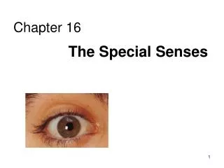
Chapter 16 The Special Senses
The Special Senses • Chemical senses • Taste (gustation) • Smell (olfaction) • Vision • The ear • Hearing • Equilibrium
Touch The sense of touch is part of the General somatic senses____ This chapter deals with the Special category of the two left sensory boxes
TASTE • Taste buds: mostly on tongue • Two types • Fungiform papillae (small, on entire surface of tongue) • Circumvallate papillae (inverted “V” near back of tongue)
Taste buds of 50-100 epithelial cells each • Taste receptor cells (gustatory cells) • Microvilli through pore, bathed in saliva • Dissolved molecules bind & induce receptor cells to generate impulses in sensory nerve fibers
Types of taste • Sweet • Sour • Salty • Bitter • Umami “beef taste”- elicited by Glutamine • Gustatory (taste) pathway to brainstem & cerebral cortex via two cranial nerves: • VII (Facial n.) – anterior 2/3 of tongue • IX (Glossopharyngeal n.) – posterior 1/3 tongue and pharynx
Smell (olfaction) Olfactory epithelium in roof of nasal cavity • Has millions of bipolar neurons = olfactory receptor cells Only neurons undergoing replacement throughout adult life Olfactory hair (cilia) bind odor molecules • Mucus captures & dissolves odor molecules Each receptor cell has an axon - are bundled into “filaments” of olfactory nerve • Penetrate cribriform plate of ethmoid bone & enter olfactory bulb
Olfactory bulb is in forebrain • In bulb nerve axons branch and synapse with mitral cells (neurons in clusters of “glomeruli”) • Mitral cells send signals via olfactory tract Filaments of Olfactory nerve (CN I) * Olfactory bulb__ _______Olfactory tract *
Anosmia: absence of the sense of smell • Trauma • Colds or allergies producing excessive mucus • Polyps causing blockage • 1/3 are from zinc deficiency • Head injury • Aging
The Eye and Vision • Vision is the dominant sense in humans • 70% of sensory receptors in humans are in the eyes • 40% of the cerebral cortex is involved in processing visual information • The eye (or eyeball) is the visual organ • Diameter 2.5 cm (1 inch) • Only anterior 1/6 visible • Lies in bony orbit • Surrounded by a protective cushion of fat
Accessory structures of the eye • Eyebrows • Eyelids or palpebrae • Upper & lower separated by palpebral fissure • Corners: medial & lateral canthi • Eyelashes
Eyelid tarsal plates give structure • Where orbicularis oculi muscles attach (close eyes) • Levator palpebrae superioris muscle • Lifts upper lid voluntarily (inserts on tarsal plate)
Tarsal glands – modified sebaceous (oil) glands in tarsal plates • Conjunctiva - transparent mucus membrane of stratified columnar epithelium • Palpebral conjunctiva • Bulbar conjunctiva • Covers white of eye but not the cornea (transparent tissue over the iris and pupil)
Lacrimal apparatus • Responsible for tears • The fluid has mucus, antibodies and lysozyme • Lacrimal gland in orbit superolateral to eye • Tears pass out through puncta into canaliculi into sac into nasolacrimal duct • Empty into nasal cavity (sniffles)
Extraocular (extrinsic) eye muscles: 6 in # • Four are rectus muscles (straight) • Lateral rectus, medial rectus, superior rectus, and inferior rectus. • Two are oblique: superior and inferior
When Extrinsic Eye Muscles Contract • Superior oblique- eyes look out and down • Superior rectus- eyes looks up • Lateral rectus- eyes look outward • Medial rectus- eyes look inward • Inferior rectus- eyes looks down • Inferior oblique- eyes look in and up
Extraocular (extrinsic) eye muscles Cranial nerve innervations: • Lateral rectus: VI (Abducens nerve) • Medial, superior, inferior rectus & inferior oblique: III (Oculomotor nerve.) • Superior oblique: IV (Trochlear n.)
3 Layers form the external wall of the eye • (outer) Fibrous: dense connective tissue • Sclera – white of the eye • Cornea • Clear because regular alignment • Role in light bending • Avascular but DOES have pain receptors • Regenerates • (middle) Vascular: • Choroid – blood rich, dark pigmented • Ciliary body – attaches lens • Iris (colored part: see next slide) • (inner) Sensory • Retina and optic nerve
(outer layer) Fibrous: dense connective tissue • Sclera – white of the eye • Cornea • (middle) Vascular: uvea • Choroid – blood rich, has dark pigmented that prevents light scattering • Ciliary body • Muscles – control lens shape • Processes – secrete aqueous humor • Zonule (attaches lens) • Iris • (inner layer) Sensory • Retina and optic nerve
Layers of external wall of eye continued • (outer) Fibrous: dense connective tissue • Sclera – white of the eye • Cornea • (middle) Vascular: uvea • Choroid – posterior, pigmented • Ciliary body • Iris • Opening is called PUPIL: lets in light • Acts like the diaphragm of a camera lens. • Regulates the amount of light that enters by contracting or dilating to see clearly. • Dark to dim light = dilation • Bright light and close vision = contraction • (inner) Sensory • Retina
Layers of external wall of eye continued • (outer) Fibrous: dense connective tissue • Sclera – white of the eye • Cornea • (middle) Vascular: uvea • Choroid – posterior, pigmented • Ciliary body • Iris • (inner) Sensory • Retina -------will cover after the chambers and lens
Chambers and fluids (see previous pics) • Vitreous humor in posterior segment • Jellylike • Forms in embryo and lasts life-time • Anterior segment filled with aqueous humor – liquid, replaced continuously • Anterior chamber between cornea and iris • Posterior chamber between iris and lens • Glaucoma when problem with drainage resulting in increased intraocular pressure
Lens: thick, transparent biconvex disc • Changes shape for precise focusing of light on retina • Onion-like avascular fibers, increase through life • Cataract if becomes clouded Cataract below: the lens is milky and opaque, not the cornea Note lens below, but in life it is clear
The eye is an optical device: predominantly the lens (to a lesser degree, not shown here, the cornea also) Note: images are upside down and reversed from left to right, like a camera • Resting eye set for distance vision: parallel light focused on retina • Resting eye doesn’tsee near objects because divergent rays are focused behind retina • Lens accommodates (becomes rounder) so as to bend divergent rays more sharply, thereby allowing convergence on the retina
Light must be focused to a point on the retina for optimal vision • The eye is set for distance vision (over 20 ft away) • 20/20 vision- at 20 feet, you see what a normal eye would see at 20 feet (20/100- at 20, normal person would see at 100) • The lens must change shape to focus for closer objects Lens Accommodation
Retina: develops as part of the brain • 1. (outer layer) Fibrous: dense connective tissue • Sclera – white of the eye • Cornea • 2. (middle layer) Vascular: uvea • Choroid – posterior, pigmented • Ciliary body • Iris • 3.(inner layer) Sensory • Retina and optic nerve Remember the 3 layers of the external eye? Retina is 2 layers • Outer thin pigmented layer: • Melanocytes (prevent light scattering) • Inner thicker neural layer • Plays a direct role in vision • Three type of neurons: • Photoreceptors • Bipolor cells • Ganglion cells
Light passes through pupil in iris, through vitreous humor, through axons, ganglion cells and bipolar cells, to photoreceptors next to pigmented layer
Photoreceptor neurons signal bipolar cells, which signal ganglion cells to generate (or not) action potentials: axons run on internal surface to optic nerve which runs to brain *Know that axons from the retina form the optic nerve, CN II
Photoreceptors: 2 types • Rod cells • More sensitive to light - vision permitted in dim light but only gray and fuzzy • Only black and white and not sharp • Cone cells • High acuity in bright light • Color vision • 3 sub-types: blue, red and green light cones *Know that rods are for B & W and cones are for color
http://www.yorku.ca/eye/rod-cone.gif http://www.secretbeyondmatter.com/ourbrains/theworldinourbrains_files/11-1.jpg
There are three types of cones • Different cones are sensitive to different wavelengths - red- long - green- medium - blue- short • Color blindness is the result of lack of one or more cone type Cone Sensitivity
COLORBLINDNESS • Comes from a lack of one or more types of color receptors. • Most are green or red or both and that is due to a lack of red receptors. • Another possibility is to have the color receptors missing entirely, which would result in black and white vision. • An inherited trait that is transferred on the sex chromosomes (23rd pair)- sex-linked trait • Occurs more often in males • Can not be cured or corrected
One of the Ishihara charts for color blindness Commonly X-linked recessive: 8% males and 0.4% females
Retina through ophthalmoscope • Macula: at posterior pole • Fovea: maximal visual acuity (most concentrated cones) • Optic disc: optic nerve exits • Vessels
Images Formed on the Retina If the image is focused at the spot where the optic disk is located, nothing will be seen. This is known as the blind spot. There are no photoreceptors there, as nerves and blood vessels pass through this point.
Green is area seen by both eyes, and is the area of stereoscopic vision Visual pathways At optic chiasm, medial fibers from each eye (which view lateral fields of vision) cross to opposite side of the brain. Optic tracts (of crossed and uncrossed fibers, sensing opposite side of visual field of both eyes) synapse with neurons in the thalamus. These axons form the optic radiation and terminate in the primary visual cortex in the occipital lobe. Left half of visual field perceived by right cerebral cortex, and vice versa.
Photoreceptors of the retina • Optic nerve • Optic nerve crosses at the optic chiasma • Optic tract • Thalamus • Visual Cortex of Occipital Lobe Visual Pathway
Visual field defectsprint this out and follow from the fields to the visual cortex using 4 colors remember: fields are reversed and upside down Visual fields Location of lesion: 1. Optic nerve ipsilateral (same side) blind eye 2. Chiasmatic (pituitary tumors classically) lateral half of both eyes gone 3. Optic tract opposite half of visual field gone 4. & 5. Distal to geniculate ganglion of thalamus: homonymous superior field (4) or homonymous inferior field (5) defect 1. 2. 1. 3. 3. 2. 4. 5. 5. 4. Visual cortex
Double vision: diplopia (what the patient experiences) • Eyes do not look at the same point in the visual field • Misalignment: strabismus (what is observed when shine a light: not reflected in the same place on both eyes) – can be a cause of diplopia • Cross eyed • Gaze & movements not conjugate (together) • Medial or lateral, fixed or not • Many causes • Weakness or paralysis of extrinsic muscle of eye • Surgical correction necessary • Oculomotor nerve problem, other problems • Lazy eye: amblyopia • Cover/uncover test at 5 yo • If don’t patch good eye by 6, brain ignores lazy eye and visual pathway degenerates: eye functionally blind NOTE: some neurological development and connections have a window of time - need stimuli to develop, or ability lost
Illusions Geometrical illusions
Illusions Successive contrast : afterimages ... fixate the black dot in the center for 60 seconds ... … and then look at a the black dot in the right panel ! what do you see?
Terminology, remember… • Optic – refers to the eye • Otic – refers to the ear • Getting eyedrops and ear drops mixed up is probably not a good idea