Download
1 / 42
470 likes | 1.38k Views
Pathology of the gall bladder and biliary tree. Pathology of the gall bladder and biliary tree. Ultrasound is considered an important investigating method in suspected gallbladder and biliary duct disease. Cholelithiasis. Presence of stones inside the gallbladder Causes :-

E N D
Pathology of the gall bladder and biliary tree Ultrasound is considered an important investigating method in suspected gallbladder and biliary duct disease.
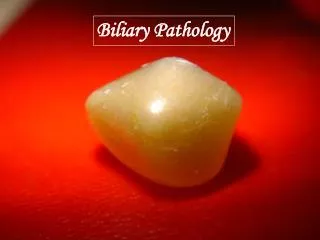
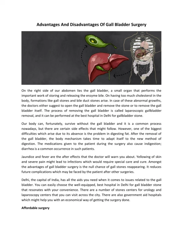
Cholelithiasis Presence of stones inside the gallbladder Causes :- • Increase secretion of bilirubin in the bile ( cirrhosis ) • Increased cholesterol in the bile occur with :- 1- Obesity 2- Diabetes 3- Pregnancy 4- Estrogen therapy • The incidence of stones rises with age because the bile flow slows down .
Cholelithiasis Ultrasound appearance :- Three classic properties of stones in gall bladder 1- Highly reflective 2- Mobile 3- Cast a distal acoustic shadow
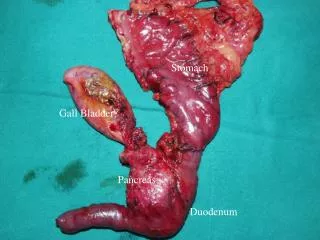
Choledocholihiasis Stones inside the CBD Stones may result from:- 1- Passage of gallstones into CBD 2- Develop de novo within the common duct.
Choledocholihiasis Clinical picture :- Often asymptomatic RUQ pain Jundice depending on the degree of obstruction Fluctuating fever if infection is present
Choledocholithiasis Ultrasound appearance:- Stone in the CBD The duct may be dilated fill or empty of stones Stones in the gallbladder Note :- Ductal stones are often not detected with US because of shadowing from duodenum.
mild duct dilatation and stone distally Ductal dilatation and stone distally
Cholecystitis • Acute or chronic Two types of acute cholecystitis :- 1- Acute calculous cholecystitis 2- Acute acalculous cholecystitis
Note :- Diffuse and marked wall thickening can also be seen in ascites, pancreatitis, hepatitis, CHF.
Gallbladder polyps • They are reflective structures project into the GB lumen • Precancerous Clinical presentation :- • Asymptomatic ( incidental finding) • Biliary colic
Gallbladder polyps Ultrasound appearance :- • Highly reflective structure • No acoustic shadow • Not movable as it has a stalk , so remain fixed with turning the patient
How can you differentiate between gall bladder stone and gall bladder polyps ?
Contracted ( small gallbladder ) Causes :- Physiological :- Postprandial Pathological :- Stones is the most common cause Microgallbladder
Contracted ( small gallbladder ) How can you differentiate between empty ( physiological small ) gall bladder and contracted one ?
Enlarged gallbladder Enlrged gallbladder refers to hydropic Causes :- Normal variant Associated with other diseases as diabetes Obstruction of cystic duct Note :- A pathologically dilated gallbladder usually assumes a more rounded appearance
Jaundice • Jaundice is a yellowish discoloration of the skin , and the sclerae (whites of the eyes), and other mucous membranes • Jaundice itself is not a disease • Caused by hyperbilirubinemia
Jaundice Jaundice is classified into three categories The three categories are:- 1- Prehepatic :- the pathology is prior to the liver 2- Hepatic: the pathology within the liver 3- Posthepatic ( obstructive ):- interruption to the drainage of bile in the biliary system
Obstructive jaundice and biliary duct dilatation Clinical presentation:- • Upper abdominal pain • May be yellow tinge of the sclera, eye and skin • Dark urine • Pale stool
Obstructive jaundice and biliary duct dilatation • Dilatation of all or part of biliary tree is usually the result of obstruction
Obstructive jaundice and biliary duct dilatation Common causes of obstruction are:- • Stones in the CBD • Neoplasm of CBD • Neoplasm of pancreatic head • liver flukes ( a group of parasite live in CBD ) less common causes of obstruction are:- • Stricture of CBD • Biliary atresia • Pancreatits
Obstructive jaundice and biliary duct dilatation Role of ultrasound in jundice :- • Differentiate between non obstructive and obstructive jaundice , by showing the dilatation of the biliary system. • The exact cause of jaundice may be difficult to identify .
Obstructive jaundice and biliary duct dilatation Notes :- • To detect the pathologically dilated gallbladder , you should look at both , the size and the shape • The dilated gallbladder has a rounded , bulging shape because of increase the pressure inside • In cases of chronic cholecystitis , the wall may be fibrotic due to stone impaction so the gallbladder lose its ability to distend , thus in these cases the Gb within normal size with dilation of biliary tree
Shotgun Sign dilated bile duct (red arrow) anterior to the portal vein (red arrowhead) resembling a double-barrel shotgun.