Summary
C linical. E ngineering. Royal Liverpool University Hospital. Comparing the Multifocal Electroretinogram response to ‘slow’ and ‘fast’ stimulation using a Roland Retiscan Richard Hagan, Anthony Fisher, Malcolm Brown

Summary
E N D
Presentation Transcript
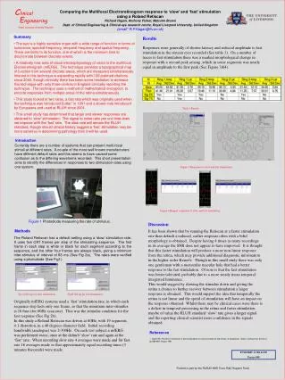
Clinical Engineering Royal Liverpool University Hospital Comparing the Multifocal Electroretinogram response to ‘slow’ and ‘fast’ stimulation using a Roland Retiscan Richard Hagan, Anthony Fisher, Malcolm Brown Dept. of Clinical Engineering & Clinical eye research centre, Royal Liverpool University, United Kingdom (email* R.P.Hagan@liv.ac.uk) • Summary • The eye is a highly sensitive organ with a wide range of function in terms of luminance, spectral frequency, temporal frequency and spatial frequency. There are limits to its function, one of which is the minimum time to discriminate between discrete events. • A relatively new area of visual electrophysiology of vision is the multifocal Electroretinogram (mfERG). This technique provides a topographical map of function from several discrete areas, which are measured simultaneously. Interest in this technique is expanding rapidly with 136 pubmed citations since 2000, though clinically there has been some hesitation to embrace this technique with only three centres in England clinically reporting the technique. The technique uses a method of mathematical encryption to encode responses from multiple areas of the retina simultaneously. • This study looked at two rates, a fast rate which was originally used when the technique was introduced Sutter1 in 1991 and a slower rate introduced by Europeans and used at RLUH since 2001. • This small study has determined that larger and slower responses are obtained to ‘slow’ stimulation. The signal to noise ratio per unit time does not improve with the ‘fast’ rate. The slow rate will remain the RLUH standard, though should clinical history suggest a ‘fast’ stimulation may be more sensitive in determining pathology then it will be used. Results Responses were generally of shorter latency and reduced amplitude to fast stimulation in the sixteen eyes recorded (See table 1). On a number of traces to fast stimulation there was a marked morphological change in response with a second peak arising ,which in some segments was nearly equal in amplitude to the first peak (See Figure 3&4). Table 1 Results Introduction Currently there are a number of systems that can present multi-focal stimuli at different rates. A couple of the more well known manufacturers have different default rates and this seems to have caused some confusion as to the differing waveforms recorded. This short presentation aims to identify the differences in responses to two stimulation rates using one system. Figure 3 Responses to slow and fast stimulation. Figure 4 Ringed responses to slow and fast stimulation. Figure 1 Photodiode measuring the rate of stimulus. Discussion Methods It has been shown that by running the Retiscan at a faster stimulation rate than default a reduced, earlier response often with a bifid morphology is obtained. Despite having 4 times as many recordings in its average the SNR does not appear to have improved. It is thought that this faster stimulation will produce a more non-linear response from the retina, which may provide additional diagnostic information in the higher order Kernels. Though in this small study there was only one gentleman with a monocular macular hole that had a better response to the fast stimulation. Of note is that the fast stimulation was better tolerated, probably due to a more steady mean temporal integrated luminance. This would suggest by slowing the stimulus down and giving the retina a chance to further recover between stimulation a larger response is obtained. This would support the idea that temporally the retina is not linear and the speed of stimulation will have an impact on the response obtained. Whilst there may be clinical cases were there is a deficit in temporal processing in the retina and faster stimulation maybe of value the RLUH standard ‘slow’ rate gives a larger signal and the reporting clinical scientist more confidence in the signals obtained. The Roland Retiscan has a default setting using a ‘slow’ stimulation rate. It uses five CRT frames per step of the stimulating sequence. The first frame in each step is white or black for each segment according to the sequence, and the other four frames are always black, giving a minimum inter-stimulus of interval of 83 ms (See Fig 2a). The rates were verified using a photodiode (See Fig1). Fig 2a Set up for slow stimulation. Fig2b Set up for fast stimulation. Originally mfERG systems used a ‘fast’ stimulation rate, in which each sequence step lasts only one frame, so that the minimum inter-stimulus is 16.6ms (for 60Hz scan rate). This was the stimulus condition for the fast response (See Fig 2b). In this study a Roland Retiscan was driven at 60Hz, with 19 segments, 4:1 distortion, in a 40 degrees diameter field. Initial recording bandwidth (analogue) was 2-300Hz. On each test subject a mfERG was performed twice, once at the default ‘slow’ rate and again at the ‘fast’ rate. When recording slow rate 4 averages were made and for fast rate 16 averages made so that approximately equal recording times (3 minutes 8seconds) were made. References 1. Sutter EE, The fast m-transform: a fast computation of cross-correlations with binary m-sequences. Siam J Comput Vol. 20, No.4, pp.686-694, August 1991 07/10/2005 11:00-12:30 Poster #49 Funded in part by the RLBUH NHS Trust, R&D Support Fund