Heart Failure
Heart Failure. Inability of heart to pump blood out as rapidly as it enters Often referred to as congestive heart failure (CHF). Congestive Heart Failure. Congestion of pulmonary or systemic circulation (backward failure) Reduced output to body tissues (forward failure). Causes. Diffuse coronary

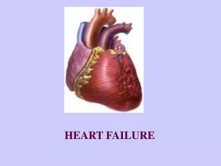
Heart Failure
E N D
Presentation Transcript
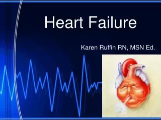
1. Heart Failure EMS Professions
Temple College
2. Heart Failure Inability of heart to pump blood out as rapidly as it enters
Often referred to as congestive heart failure (CHF)
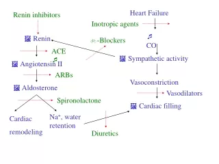
3. Congestive Heart Failure Congestion of pulmonary or systemic circulation (backward failure)
Reduced output to body tissues (forward failure)
4. Causes Diffuse coronary artery disease
Myocardial ischemia
Myocardial infarction
Arrhythmias
Tachycardia
Bradycardia
5. Causes Valvular heart disease
Acute Hypertensive Crisis
Chronic Hypertension
Idiopathic Causes
6. CHF May develop acutely or may be a chronic disease
Acute Onset CHF: Suspect
Acute MI
Dysrhythmia
Hypertensive Crisis
7. CHF Chronic CHF may worsen acutely from:
Respiratory infection
Pulmonary embolism
Emotional stress
Increased salt and water intake
8. Congestive Heart Failure Left sided
Right sided
Biventricular
9. Left-Sided Heart Failure Left ventricle fails as effective pump
Left ventricle cannot eject blood delivered from right heart through pulmonary circulation
Blood backs up into pulmonary circulation
10. Left-Sided Heart Failure Increase pressure in pulmonary capillaries forces blood serum out of capillaries into interstitial spaces and alveoli
Increase respiratory work and decrease gas exchange occur
11. Left-Sided Heart Failure Common causes
ACUTE MI
especially if involves left ventricle
Chronic hypertension
Dysrhythmias
especially tachydysrhythmias
12. Left-Sided Heart Failure Pulmonary Signs/Symptoms
13. Left Heart Failure Symptoms Dyspnea on exertion
Paroxysmal nocturnal dyspnea
Orthopnea
Fatigue, generalized weakness
14. Left Heart Failure Signs Anxiety, confusion, restlessness
Persistent cough
Pink, frothy sputum
Tachycardia
Tachypnea
Noisy, labored breathing
Rales, wheezing (�cardiac asthma�)
Cyanosis (late)
Third heart sound (S3)
15. Right-sided Heart Failure Right ventricle fails as effective pump
Right ventricle cannot eject blood returning through vena cavae
Blood backs up into systemic circulation
16. Right Heart Failure Increased pressure in systemic capillaries forces fluid out of capillaries into interstitial spaces
Tissue edema occurs
17. Right Heart Failure Causes
18. Right Heart Failure Causes Others
Chronic hypertension
COPD (cor pulmonale)
Pulmonary embolism
Right ventricular infarction
19. Right-Sided Heart Failure Systemic Signs/Symptoms
20. Right Heart Failure Signs/Symptoms Tachycardia
Jugular vein distension
Pedal, pre-tibial, sacral edema
Hepatomegaly
Splenomegaly
21. Right Heart Failure Signs/Symptoms Anasarca (generalized edema)
Fluid accumulation in body cavities
Ascites
Pleural effusion
Pericardial effusion
22. Management of Heart Failure
23. Goals of Management Improve oxygenation, ventilation
Decrease venous return to heart
Decrease cardiac work, O2 demand
Improve cardiac output by
Reducing afterload
Increasing myocardial contractility
24. Management Sit patient up, dangle feet
Do not lay flat
Oxygen by non-rebreather mask
Consider positive pressure ventilation
25. Management Consider intubation if:
O2 saturation cannot be kept >90% on 100% O2
PaO2 cannot be kept >60 torr on 100 % O2
Patient displays signs of worsening cerebral hypoxia
PaCO2 progressively increases
Patient becoming exhausted
26. Management Monitor ECG
Hypoxia, increased heart wall tension leads to dysrhythmias
IV NS TKO via microdrip or lock
Limit Fluids
If RVF only, fluid challenges to ? preload
27. CHF First Line Drug Therapy Nitroglycerin
0.4mg SL q 5 min prn
Systolic BP should be > 90 - 100 mm Hg
Nitrate therapy before IV is started
Reduces preload/afterload
Improves coronary artery perfusion
Caution in RVF
NTG, Lasix or MS may worsen hypotension
Use inotropes if fluid does not improve BP following NTG administration
28. CHF First Line Drug Therapy Furosemide (Lasix�) -
40 mg (0.5 - 1 mg/kg) slow IV
Patients already on furosemide may have tolerance
Increase dose to 2X daily oral dose
Direct vasodilation leads to decreased venous return
Diuresis leads to decreased intravascular volume
May cause hypokalemia, dysrhythmias
especially dangerous if patient on digitalis
May worsen hypotension in RVF
29. CHF First Line Drug Therapy Morphine Sulfate
2 mg IV push slowly q 10-15 min
Peripheral vasodilation leads to
Decreased preload
Decreased afterload
Decreased venous return leads to
Decreased cardiac work
Decreased O2 demand
Decreased anxiety
Decreased release of catecholamines
Monitor Ventilations and BP
Systolic BP should be > 90 - 100 mm Hg
30. CHF Second Line Therapy Dobutamine
2 - 20 mcg/kg/min
Potent ?1 stimulation
Increases contractility
Increases level of cardiac output
Drug of choice if systolic BP >100 and diastolic BP <110
31. CHF Second Line Therapy Nitroglycerin
10 mcg/min increased by 5-10 mcg/min q 5 min
Vasodilation
Decreased venous return leads to
Decreased cardiac work
Decreased O2 demand
Decreased afterload leads to increased cardiac output
32. CHF Third Line Drug Therapy Bronchodilators (beta agonists)
May be useful if wheezing is present
Mild peripheral vasodilator
Myocardial and respiratory stimulant
May cause arrhythmias in hypoxic patients or those with coronary artery disease
33. CHF Management What if the BP is too low for the first and second line drug therapies?
BP < 70 mm Hg
norepinephrine, 0.5 - 30 mcg/min IV infusion
BP > 70 but < 100 mm Hg
dopamine, 5 - 15 mcg/kg/min IV infusion
After BP improves, treat pulmonary edema with first and second line therapies
34. CHF Management Long Term Management usually includes
Fluid minimization
Diuretics (+ Potassium if non-potassium sparing)
Diet restrictions
Increase contractility
Digitalis
Blood pressure control
ACE Inhibitors
Coronary artery perfusion
Nitroglycerin
35. Cardiogenic Shock
36. Cardiogenic Shock Diminished cardiac output leading to impaired tissue perfusion
Most extreme form of pump failure
37. Cardiogenic Shock Occurs in about 15% of acute MI patients
Usually occurs when 40% or more of the left ventricular muscle mass infarcts
Mortality is 85% or more with treatment
38. Signs/Symptoms Confusion, restlessness, anxiety, stupor, coma
Cool, clammy skin
Pallor
Weak or absent extremity pulses
Tachycardia
Slow or absent capillary refill
39. Signs/Symptoms BP < 90 systolic or > 30mmHg below normal
BP is NOT the same as perfusion
Shock can be present with a �normal� BP
Evaluate signs of peripheral perfusion in addition to BP
40. Cardiogenic Shock Very difficult to assess in presence of arrhythmias, hypovolemia, decreased vascular tone
41. Cardiogenic Shock Treatment Priorities:
Rate
Rhythm
BP (Volume, Pump/Vascular tone)
Correct major disorders of rate, rhythm before directly treating BP
42. Goals of Management Improve oxygenation and peripheral perfusion
Avoid increasing cardiac workload
myocardial oxygen demand
43. Management Primary assessment & Focused Hx
Identify source of problem
Acute pulmonary edema
Volume problem
Pump problem
Rate problem
44. Acute Pulmonary Edema First line interventions
IV/O2/ECG Monitor
If BP > 90-100 mm Hg:
furosemide 0.5 � 1.0 mg/kg slow IV (or twice patient�s single daily dose up to 120 mg)
Morphine 2 � 10 mg slow IV
Nitroglycerin 0.4 mg SL
If BP < 90 mm Hg:
Vasopressors based on SBP
45. Volume Problem IV/O2/ECG Monitor
Fluid challenge until rales or if evidence of anterior wall AMI
Vasopressors based on SBP
46. Pump Problem IV/O2/ECG Monitor
SBP <70 mmHg:
norepinephrine 0.5 � 30 mcg/min IV inf
SBP 70 � 100 mm Hg & shock
dopamine 5 � 15 mcg/kg/min IV inf
SBP > 100 mm Hg w/o shock
dobutamine 2 � 20 mcg/kg/min IV inf
47. Management Keep patient supine
Difficult in presence of pulm edema
Do not elevate lower extremities
Oxygenate via NRB
Consider assisting ventilations
Decrease work of breathing may benefit patient in shock
Consider intubation
Monitor ECG
48. Management IV TKO with microdrip set or lock
Limit fluids unless suspect RVF
Correct major disorders of rate, rhythm
Increase rate in bradycardias
Terminate tachycardias with cardioversion
Suppress frequent ectopic beats
49. Management If rate/rhythm adequate, treat BP
Consider fluid challenge of 250cc LR over 10-15 minutes if relative or absolute hypovolemia possible, including RVF and NO pulmonary edema
Avoid use of vasopressors until volume deficits corrected or pulmonary edema presents
50. BP Treatment Review If rate, rhythm, volume adequate, treat BP with vasopressors:
Norepinephrine, or
Dopamine
51. Norepinephrine 0.5 - 30 mcg/min
Inotropic and vasoconstrictive properties
Can be used if systolic BP < 70
If systolic BP > 70, use dopamine instead
DO NOT use until hypovolemia corrected
DO NOT allow infiltration
52. Dopamine 2 - 20 mcg/kg/min
Place 200 mg/250cc of D5W
Begin at 5 mcg/kg/min
In 2 - 10 mcg/kg/min range, ? effects dominate
> 20 mcg/kg/min ? effects dominate
Use lowest dose that produces good perfusion
Use as initial vasopressor if BP 70-100 systolic
If dopamine infusion rate is > 20 mcg/kg/min use norepinephrine
53. Dopamine May cause tachycardia, ectopy, nausea
DO NOT use until hypovolemia is corrected
DO NOT allow to infiltrate