Download
1 / 28
280 likes | 418 Views
Visualizing the localization of protein isoforms in HeLa cells with laser confocal microscopy. Justin R. Siebert Nancy J. Bachman, Ph.D. Biology Department State University of New York College at Oneonta, Oneonta NY 13820. The Beginning.

E N D
Visualizing the localization of protein isoforms in HeLa cells with laser confocal microscopy Justin R. Siebert Nancy J. Bachman, Ph.D. Biology Department State University of New York College at Oneonta, Oneonta NY 13820
The Beginning • Neighbor of cytochrome c oxidase subunit IV (NOC4) gene shares a bidirectional promoter with subunit IV • Comparative Gene Identification isolate 112 (CGI-112) discovered using BLAST • NOC4 and CGI-112 are 40% identical in amino acid sequence
CGI –112 • CGI-112 and NOC4 comprise a novel protein family • The CGI-112 gene is located between the PA28 and subunits of the 11S proteasome regulator on chromosome 14
CGI-112 Function and Cellular Associations ?
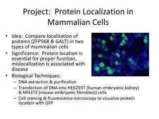
Questions • Where in the cell does CGI-112 localize? • Do CGI-112 and NOC4 proteins co-localize? • Do CGI-112 or NOC4 co-localize/interact with the proteasome? • Does the overexpression of NOC4 or CGI-112 induce apoptosis?
Earlier Studies • Earlier studies attempted back at SUNY Oneonta • Only able to view GFP • The microscope available: Zeiss standard 16 epifluorescence microscope • Imaging abilities limited • 35mm slide film • POOR QUALITY
Fall 2004 Experiment The Laser Scanning Confocal Microscope presented a new opportunity to address the questions asked in the earlier slide
Cell Culture • HeLa cells (ATCC) were seeded at about 30% confluence in 60 mm dishes • Sterile, uncoated, #1.5 square coverslips placed in well • Cultures grown in Dulbecco's Modified Eagles Medium + 10% fetal calf serum + 0.5 mM non-essential amino acids • Cultures were incubated at 37◦ C in 5% CO2 for about 20 hrs
Transfection • Plates were incubated with 1.6 g plasmid DNA containing either human retinal NOC4-GFP or human colon CGI-112-GFP complexed with Lipofectamine and Plus Reagent. • Transfected plates were supplemented with fresh plating medium after 4 hrs, then cells were incubated for about 16 hrs at 37◦ C in 5% CO2.
Antibody Labeling • Plates were washed 3x in 1x Phosphate Buffered Saline (PBS). • Cells were fixed in 4% paraformaldehyde for 10 min. at room temperature. • Plates were washed 3x in 1x PBS, then permeabilized in 1x PBS/0.2% Triton X-100 and incubated 30 min. at room temperature.
Antibody Labeling • Plates were rinsed once in PBS, then incubated in 10% BSA for 30 min. at room temperature. • Plates were washed 3x, in 1x PBS. • Primary antibodies (Rabbit anti-NOC4 or Rabbit anti-PA28α) were diluted 1:200 in 1% BSA in PBS.
Antibody Labeling • Plates were incubated with appropriate antibody dilutions for 2 hours at room T. • Plates were washed 5x in 1x PBS, then incubated in a 1:1000 dilution of Texas Red-X conjugated goat anti-rabbit IgG in 1% BSA in PBS. • Plates were washed 3x in 1x PBS. Coverslips were rinsed in ddH2O and mounted separately on glass slides in mounting medium or Vectashield mounting medium with DAPI.
Laser Confocal Microscopy Slides were visualized on the Zeiss LSM 510 META at Binghamton University Slides were viewed 1 week after preparation at SUNY Oneonta Slides were kept in the dark, and in refrigerator until viewing DAPI, Texas Red-X, and GFP, were fluorochromes used to mark cellular components
DAPI • DAPI will stain the nuclear material in the cell; designated color is blue. • Source: Vectashield mounting media. Should stain every HeLa cell nucleus • Laser: Violet/Blue laser diode (405nm diode), using the DAPI filter setting on the microscope. • The DAPI stain provides internal controls • shows HeLa cells are not autofluorescent, • shows other dyes do not bind at random to cells/cellular components.
Texas Red-X Texas Red-X will be used to detect binding of specific primary antibodies (PA28a or NOC4). Color designated for TR is red. Source: secondary antibody labeling, Texas Red-X conjugated to goat anti-rabbit IgG. Laser: HeNe 543 Laser, using the CY3 filter setting on the microscope ONLY cells marked with the primary antibody should stain red
GFP GFP is fused in-frame at the carboxy terminus of each of the two proteins. Color designation for GFP is green. Source: Plasmid DNA for NOC4-GFP and CGI-112-GFP fusion proteins Laser: Argon Ion Laser (488nm), using the FITC filter on the microscope. GFP should appear only in transfected cells. When viewed, the proteins NOC4-GFP and CGI 112-GFP should show up green.
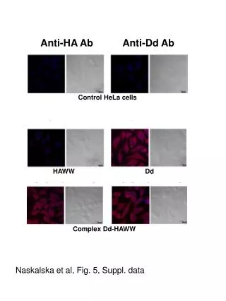
NOC4/ PA28a Blue – DAPI Red – Texas Red PA28a Green – GFP NOC4 Yellow- Colocalization 1.0 Zoom, 40x Oil Objective, Multi track picture, DAPI, FITC, CY3 channels activated.
NOC4/ PA28a Blue – DAPI Red – Texas Red PA28a Green – GFP NOC4 Colocalization 1.0 Zoom, 40x Oil Objective, Multi track picture, DAPI, FITC, CY3 channels activated.
PA28a / CGI-112 Blue – DAPI Red – Texas Red PA28a Green – GFP CGI-112 Yellow- Colocalization 1.0 Zoom, 40x Oil Objective, Multi track picture, DAPI, FITC, CY3 channels activated.
PA28a / CGI-112 Blue – DAPI Red – Texas Red PA28a Green – GFP CGI-112 Yellow- Colocalization 1.0 Zoom, 40x Oil Objective, Multi track picture, DAPI, FITC, CY3 channels activated.
NOC4 / CGI-112 Blue – DAPI Red – Texas Red NOC4 Green – GFP CGI-112 Yellow- Colocalization 1.0 Zoom, 40x Oil Objective, Multi track picture, DAPI, FITC, CY3 channels activated.
Initial Conclusions It appears that expression of transfected CGI-112 or NOC4 is necessary for binding of PA28a antibody. The NOC4 and CGI-112 proteins mostly colocalize. The NOC4 and CGI-112 proteins mostly colocalize with the PA28a subunit of the 11S proteasome regulator.
New Questions • What are the “cytoplasmic holes” found in the transfected cells? • Are the transfected HeLa cells undergoing apoptosis? 2.0 Zoom, 40x Oil Objective, Multi track picture, FITC, CY3 channels activated. Red – Texas Red - PA28a Green – GFP - NOC4 Yellow- Colocalization Slide showing NOC4/PA28a. Mysterious “holes” observed
New Questions • In the experiments looking at colocalization of PA28a and CGI-112 why are there some green dots, and yellow dots? 2.0 Zoom, 40x Oil Objective, Multi track picture, DAPI, FITC, CY3 channels activated. 2.3 Zoom, 40x Oil Objective, Multi track picture, FITC, CY3 channels activated. C
Acknowledgements Dr. Dennis McGee for microscope training Binghamton University LSCM facility