Download

1 / 23
290 likes | 784 Views
Electrolytes. Clinical Pathology. Electrolytes. Electrolytes and acid-base disorders may result from many different diseases. Correction of fluid, electrolytes, and acid-base disturbances is often more immediate benefit to patients than a specific diagnosis.

E N D
Electrolytes Clinical Pathology
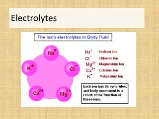
Electrolytes • Electrolytes and acid-base disorders may result from many different diseases. • Correction of fluid, electrolytes, and acid-base disturbances is often more immediate benefit to patients than a specific diagnosis. • Most common electrolytes that are measured are Na+, K+, Cl-, and HCO3, as TCO2. • Serum is the best place to observe the electrolyte levels.
Anion Gap • Used to determine metabolic acidosis • (Na++K+)- (Cl- + HCO3)
Sodium • Most abundant electrolyte in blood. • Functions: • Maintain osmotic pressure • Acid-base balance • Transmit nerve impulses • Essential for renal water retention (controls hydration status).
Hyponatremia • Diabetes Mellitus (DM) • Addison’s disease • Diarrhea (foals and horses) • Renal disease (cattle) • Salt deficiency (cattle) • Ruptured urinary bladder (horse, dog, cat) • Saliva loss (horse) • Psychogenic polydipsia
Hypernatremia • Panting • Sweating • Diabetes insipidus • Increased GI water in ruminants (grain overload acidosis, propylene glycol toxicity).
Normal Sodium Values • Normal values: • Dogs: 140-150 mEq/L • Cats: 150-160 mEq/L Neurologic signs may occur at <120 or >170 mEq/L in dogs.
Serum Chloride • Important in many secretions • Saliva • Sweat • gastric • Increases and decreases may parallel change in serum sodium • Normal values: • Dogs: 105-115 mEq/L • Cats: 115-125 mEq/L Danger values are unknown
Potassium • Serum potassium is maintained within narrow limits for normal neuromuscular and cardiac function. • Potassium is released from platelets during clotting. • Normal values: • Dogs: 3.5-5.5 mEq/L • Cats: 3.5-5.5 mEq/L Danger values are <2.5 mEq/L May result in cardiac conduction disturbances
Hyperkalemia and Hypokalemia • Hyperkalemia • Anuria • Addison’s • Parenteral administration • Hypokalemia • Loss through GI fluids, urine or anorexia.
Calcium • Dietary intake rarely effects serum levels directly • 99% of Calcium is stored in the bone, other in cells and extracellular fluid • Functions: • Main component of bones and teeth • Cofactor for clotting • Necessary for transmission of nerve impulses and muscle contraction
Hypercalcemia and Hypocalcemia • Hypercalcemia • Renal failure (horses) • Neoplasia • Certain plants • Addison’s • Hypocalcemia • Hypoproteinemia • Milk fever (eclampsia) • Hypomangesmic tetany • Panceatitis
Blood Gas Anaylsis • Useful in any severely ill dog or cat (vomiting, diarrhea, etc) • Analysis of proper evaluation of gas exchange and alterations of TCO2 in patients with respiratory disorders • Analyzers are equipped with specific electrodes to measure pH carbon dioxide tension (pCO2), and oxygen tension (pO2). • Arterial blood is ideal but jugular vein blood may be used.
Blood Gas Analysis Continued • Blood is collected in a heparinized syringe. • The blood is immediately injected into the machine for analysis • Test takes 15-30 minutes • Used to determine if animal is in metabolic acidosis/alkalosis or respiratory acidosis/alkalosis.
Metabolic Acidosis • Characterized by decreased plasma HCO3, decrease pH, and decrease pCO2 • Loss of HCO3 usually occurs via the GI tract but may also occur via the kidneys
Respiratory Acidosis • Due to decreased effective ventilation (increased pCO2). • Decrease pH and compensatory increase in HCO3. • Hypoventilation may occur from airway obstruction, cardiopulmonary arrest, and neuromuscular diseases.
Metabolic Alkalosis • Increased plasma HCO3, increased pH, and compensatory increased CO2. • Caused by loss of chloride rich fluid via the GI tract
Respiratory Alkalosis • Results from increased ventilation. • Decreased pCO2, increased pH and decrease HCO3. • Caused by tachypnea due to hypoxemia usually secondary to a disease process.