Positive Electrolytes
450 likes | 501 Views
Explore electrolyte concentrations in body compartments, conversions, sodium control, potassium balance, and related disorders.

Positive Electrolytes
E N D
Presentation Transcript
3rdYear Inorganic Pharmaceutical Chemistry, Pharmacy CollegeBody Major ElectrolytesPositive and Negative Ions Dr. Kassim Mohammed
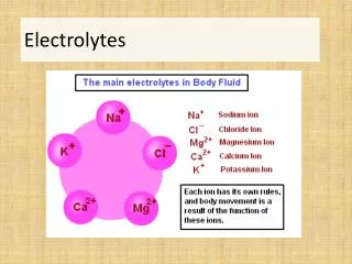
The Body Compartments The electrolyte concentration will vary with a compartment. The three body fluid compartments are; 1. Intracellular fluid (45-50%) of body weight. 2. Extracellular fluid, made of two parts; A) Interstitial fluid (12-15%) of body weight. B) Vascular fluid or plasma ( 4-5%) of body weight. The three compartments are separated from each other by permeable membranes. The later allows the passage of water and some inorganic as well as organic substances.
Units of Concentration Electrolyte concentrations are expressed by units of mEq/l or w/w %. Eq.wt ÷ mEq/l = mg/l Eq./mole ÷ Mol. wt = Eq.wt Ex. Calculate the amount of salt necessary to make a solution that contains 153mEq/l each of Na or chloride ions and state w/w %
Ex2: What is the weight of CaCl2 .2H2O needed to prepare a liter of solution containing Ca+29mEq / l? atomic masses for Ca and Cl are 40 and 35.5 respectively. Ex3: What will be the chloride content of a solution containing 661.5 mg CaCl2 .2H2O?
SODIUM Sodium is the principal cation in extracellular fluid. Its responsible for maintaining osmotic pressure and hydration. Its absorbed from daily diet by the intestinal tract. Excess sodium is excreted by the kidneys, approximately 80-85% of sodium is reabsorbed. A complex hormone system may be involved in the reabsorption of sodium. Renin, angiotension I and II and aldosterone –produced by the adrenal cortex-which regulates the absorption of sodium in renal tubules.
Hyponatremia Conditions causing hyponatremia (low sodium serum level) include: 1. Extreme urine loss such as seen in diabetes insipidus which caused by deficient insulin secretion by the beta cells of the islets of Langerans in the pancreas. 2. Metabolic acidosis, in which sodium is excreted. 3. Addison’s disease with decreased excretion of the ADH, aldesterrone . 4. Diarrhea and vomiting 5. Kidney damage
Hypernateria Hypernateria may be caused by: 1. Cushing’s syndrome with increased in ADH, aldesterone production, 2. Severe dehydration, 3. Certain types of brain injury, 4. Excess treatment with sodium salts.
Sodium level and Hypertension Sometimes the body is unable to eliminate sodium and the concentration starts to increase, water is retained in the tissues to maintain osmotic balance. Edema results and the patient can take a puffy appearance with swelling, particularly of the lower extremities. The buildup of fluids puts an added burden on the heart which may be aggravated if the heart is also diseased. Treatment includes low salt diets, diuretics, cardiotonic drugs or combination of each.
Sodium Control and Replacement Sodium – free salt substitutes can be used to enhance the flavor of food. A wide variety of these are now available in the market such as Neucartasal and Co-Salt mixtures. Sodium Chloride: Oral 1 gram three times a day or IV 1 liter of a 0.9% solution. Fructose and sodium chloride injections; 10% fructose and 0.9% NaCl. It is nutrient and electrolyte replenisher.
POTASSIUM Potassium is the major intracellular cation present in concentrations approximately 23 times higher than the concentration of potassium in the extracellular fluid. The small fraction 2% of total body potassium which is in the extracellular fluid is distributed proportionately between the interstitial and the plasma. The concentration in serum is around 4.5mmol/l. The concentration content is maintained by an active transport mechanism. During transmission of a nerve impulse, potassium leaves the cell and sodium enters the cell, sodium-potassium pump.
Potassium in the diet is rapidly absorbed and the excess potassium is rapidly excreted by the kidneys. Potassium salts have been used for their diuretic action because of the efficient excretion of potassium by the kidneys, since a certain volume of urine will be excreted in order to keep the potassium salt in solution. Whole body counts of potassium can be found by measuring levels of potassium - 40.
Hypopotassemia Hypopotassemia can be serious to the patient. It causes changes ECG and in myocardial function, flaccid and feeble muscles and low blood pressure. The main causes of hypopotassemia are; 1. Vomiting and Diarrhea 3. Burns 4. Hemorrhage 5. Diabetic coma 6. IV infusion of solution lacking in potassium 7. Overuse of thiazide diuretics 8. Alkalosis, movement of potassium into cells as protons move out of the cell into the proton deficient extracellular fluid.
Hyperpotassemia Hyperpotassemia is less common and occurs during certain types of kidney damage. If the kidney is functioning properly the body can eliminate excess potassium readily. In certain acidotic conditions, interference with the sodium and potassium proton exchange can result in potassium retention. Potassium may be released from some damaged cells leading to increases serum potassium. Potassium Replacement 1. Potassium chloride, irritant to gastrointestinal tract. 2. Potassium Gluconate, less irritating than the chloride.
Potassium Level and the Heart The heart is sensitive to potassium concentrations. In hypopotassemia there are alterations in the ECG (fattened T wave) and disticthitological alterations in the myocardium. Hyperpotassimea also results in changes in the ECG (peaked T wave) and causes the heart muscles to become flaccid with possible cessation of heart beat (potassium unrest). It is thought that potassium may replace calcium in the cardiac muscle since a decrease in calcium will produce a similar pattern in heart muscle and may explain why calcium glutamate is effective in hyperpotassimia conditions.
CALCIUM About 99% of body potassium is found in bones and the remaining is in ECF. Calcium is absorbed by the upper part of the intestinal track where the contents are still acidic. At neutral or alkaline media calcium is precipitated as the dibasic phosphate CaHPO4, carbonate, oxalate and sulfate salts and as insoluble calcium soaps. The fatty acid portion of the soaps comes from lipase – catalysed hydrolysis of dietary triglycerides.
Calcium Absorption Calcium absorption across the intestinal walls is controlled by the parathyroid hormone, PTH, and a metabolite of vitamin D3. The activated metabolite, 1,25-dihydroxycholecalciferol, may function as a gene activator causing the synthesis of calcium binding protein which transfer the calcium ions across the intestinal walls. Epileptic children on anticonvulsant may have low calcium levels.
Phosphates concentration affects the intestinal absorption and serum calcium level. Increased serum phosphorus level will lower serum calcium. The administration of phosphorus salts has been used with some success in the treatment of hypercalcemia.. Lactose and Calcium There is also evidence that lactose plays a role in calcium absorption with lactose-deficient patients having a higher incidence of osteoporosis.
Blood Ca ions Level and the PTH Blood calcium levels control the secretary activity ِthe parathyroid gland: decreased blood calcium increases parathyroid secretion and vice versa. Removal of the this gland will lead to muscle tetany as a result of severe drop in calcium levels and the rise in phosphate levels. PTH controls both calcium and phosphate levels by acting on the kidneys and the bone. Administration of PTH raises the blood calcium and decreases the blood phosphate.
The hormone calcitonin also affects calcium absorption. Its action on bone is to inhibit calcium resorption. In the kidneys calcitonin increase the urinary excretion of phosphate by an indirect effect. Because calcitonin produces hypocalcemia, PTH is released causing urinary phosphate excretion.
99% of the body calcium is found in bone, as hydroxyapatite. The remaining ionic calcium is involved in the neurohormonal functions., blood clotting, muscle contraction and other biochemical processes. Calcium is necessary for the release of acetylcholine from nerve endings. Muscles become flaccid when calcium is removed or displaced. Heart muscles are affected when potassium displaces calcium in hyperpotassemia.
Another main role of calcium in body is in blood clotting. This can be avoided when citrate is added to complex calcium hence preventing clot formation in the collected blood. Treatment is urgent if the serum calcium is greater than 3.5 mmol/l. IV saline is administered to restore the glomularfilteration rate and promote diuresis. Steroids, calcitonin and IV phosphate have been used to lower calcium concentration. Biophosphate and aminohydroxyprpoylidene have been proved to be the best in lowering serum calcium. Surgical removal of a parathyroid adenoma usually provides a complete cure. Immediately after successful surgery transient hypocalcaemia may have to be treated with vitamin metabolites.
Symptoms of hypercalcaemia fatigue, muscle weakness, constipation, anorexia and cardiac irregularities. If the conditions persists calcium salts may deposit in kidneys and blood vessels. Methods of reducing intestinal calcium absorption include; 1. Precipitation of calcium as insoluble calcium sulfate pr phosphate salts. 2. Complexation with EDTA. 3. Using cellulose phosphate.
Causes of Hypercalcemia Hypercalcemia is found in; 1. Hyperparathyroidism 2. Hypervitaminosis D, e.g.in treatment of hypoparathyroidism or renal disease. 3. Bone neoplastic disease 4. Diuretic therapy, the hypercalcamea is usually mild. 5. Immobilisation: especially in young people or patients with Paget’s disease. 6. Milk alkali syndrome: the combination of increased calcium intake together with bicarbonate, as in a patient self medicating with proprietary antacid.
Hypocalcemia can be caused by; 1. Hypoparathyroidism 2. Vitamin D deficiency 3. Osteoblastric metastasis. 4. Cushing’s syndrome (hyperactive adrenal cortex) 5. Acute pancreatitis 6. Acute hyperphosphatemia.
Calcium Control When a person is fasting or sleeping reabsorption of bone takes place in order to maintain blood calcium levels. In osteoporosis, the bones become weaker and more fragile with broken hips is commonly seen in elderly with this disease. Page’s disease is another problem associated with calcium metabolism. This may be treated using phosphate salts and or calcitonin. Calcium Replacement: 1. Calcium chloride contains 0.033% CaCl2 .2H2O 2. Calcium gluconate
Magnesium After potassium, magnesium is the second most prevalent cation in ICF and the fourth most abundant cation in the body. About 50% of magnesium is in bones. It is also essential in protein synthesis and for the functioning of neuromuscular system. Electrical properties of cell membranes are affected by any reduction in extracellular magnesium concentration. Some 300 enzyme systems are magnesium dependant.
Magnesium influences the secretion of PTH by the parathyroid glands. It affects glycolysis, oxidative metabolism and transmembrane transport of potassium and calcium. Causes of hypomagnesia include: Malnutrition Dietary restriction Chronic alcoholism Faulty absorption or utilization Gastrointestinal diseases Osmotic diuresis such as occurs in diabetes mellitus. Medications, for example treatment with immunosuppressant drug cyclosporine. Parathyroid hormone imbalances.
The repeated demonstration of a magnesium concentration of less than 0.7 mmol/l in a serum specimen is evidence of marked intracellular depletion and of clinical condition which may benefit from magnesium therapy. Normal subjects retain 90% of IV test material compared to 40% in patients with hypomagnesia. Symptoms of hypomagnesia include: Personality changes after depletion of 3-4 months. Failure to gain weight properly. Cardiac disturbances.
Magnesium ion has a definite pharmacological action which resembles that produced by chloroform. This depressant action affects the cellular portion of the neuron and the neuromuscular junction. An excess of magnesium decreases the amount of the neuro transmitter substance, acetylcholine. Calcium ions relieve the block produced by magnesium ions and restore output of acetylcholine from nerve endings. The alkalinity of the gastrointestinal tract reduces the absorption of magnesium which normally takes place at the upper part of the intestinal tract, the duodenum.
Magnesium supplements in oral diets is complicated by the fact they often cause diarrhea. A variety of oral, intramuscular and intravenous regimes have been proposed. In any case must be taken in case of impaired kidney function to avoid toxicity. Magnesium Replacement: Magnesium Sulphate, when injected used as CNS depressant, 4 grams in 10% solution. Magnesium sulphate; Oral dose 1-10 grams daily.
Chloride It’s the major extracellular anion and is responsible for maintaining osmotic pressure, proper hydration and normal cation - anion balance in the plasma and interstitial fluid compartments. Chloride ions are absorbed from food in the intestinal tract and is removed from blood by glomerular filtration and possibly reabsorbed by the kidney’s tubules. The chloride ions, as such, has no particular pharmacological activity.
Hyporchloremia Hypochloremia can be caused by; Salt – losing nephritis ( inflammation of the kidney). Metabolic acidosis as in diabetes mellitus and renal failure, leading either to excessive production or diminished excretion of acids leading to the replacement of chloride by acetoacetate and phosphate. Prolonged vomiting with loss of chloride as gastric hydrochloric acid. Hyperchloremia can be caused by; Dehydration decreased renal blood flow found with congestive heart failure, severe renal damage excessive chloride intake.
PHOSPHATE Phosphate is abundant in the body and is an important ICF and ECF anion. Much of the phosphate in the body is attached to lipid and proteins. Most of the body phosphate is in bone. Phosphate changes accompany calcium deposition or reabsorption by bone. Control of ECF phosphate is achieved by the kidney, where tubular reabsorption I reduced by PTH. The phosphate which is not reabsorbed in the renal tuble acts as an important urinary buffer.
PHOSPHATE….continued Most phosphate salts of pharmaceutical concern and are phosphate esters. Their biochemical interest are derived from phosphoric acid, commonly written as H3PO4 but more accurately represented as PO(OH)3. This acid is also known as orthophosphoric acid. Other important phosphate forms are metaphosphoric acid and pyrophosphoric acid. Common phosphate salts of pharmaceutical importance are sodium dihydrogen phosphate, sodium monohydrogen phosphate and sodium phosphate. In ECF the total concentration of both monohydrogen phosphate and dihydrogen phosphate is maintained in the limits 0.8-1.4 mmol/l. This phosphate must be distinguished from organically bound phosphate such as in ATP.
PHOSPHATE….continued The main phosphate ion in intracellular fluid compartment is HPO4-2 Its main role can be summerised a follows: 1. ATP is the potential chemical energy storage which contains the phosphoric acid anhydride linkage. 2. The phosphate is important in the buffer system HPO4-2/ H2PO4- 3. The sugars hexoses are metabolized as phosphate esters.
PHOSPHATE….continued 4. Phosphorous is essential for the proper calcium metabolism. 5. Phosphorous is essential for normal bone and tooth development since it is a component in hydroxyapatite, the main calcium salt found in bone and teeth. Serum Phosphate and Calcium Levels There is a correlation between serum phosphate levels and calcium levels. Whenever calcium concentrations are not within normal range, serum phosphate will either be too high or too low. In plasma, there is a reciprocal relationship between calcium and phosphate.
Hyperphosphatemia may be caused by; 1. Hypervitaminosis D increases intestinal phosphate absorption along with calcium. 2. Renal failure due to the inability to excrete phosphate into the urine, Phosphate excretion is impaired. 3. Hypoparathyroidism, the lack of parathyroid hormone permits renal tubular reabsorption of phosphate which results in decrease of urinary phosphate and a rise in serum concentration. 4. Haemolysis may occur intravascularly in the patient, or may be a consequence of an improper sampling procedure. 5. Pseudohyperparathyroidism. There is tissue resistance to PTH.
PHOSPHATE….continued Hypophsphatemia is uncommon because a balanced diet contains adequate amounts of phosphate. Only in patients on IV solutions hypophosphatemia may arise. It causes marked alterations in erythrocyte metabolism and may be seen in; 1. Vitamin D deficiency(rickets) probably caused by decreased intestinal calcium absorption. 2. Hyperparathyroidism, increased levels of parathyroid hormone further inhibit renal tubular phosphate reabsorption, resulting in increased urinary phosphate excretion, hence decreased serum phosphate levels.
PHOSPHATE….continued 3. Lack of phosphate reabsorption by kidney tubule from other causes e.g. infection and cancers. 4. Long-term aluminum hydroxide gel antacid therapy. This compound forms insoluble aluminum phosphate salts from dietary phosphate therapy hence preventing phosphate absorption from the intestinal tract. Dibasic calcium phosphate is given orally as a source of calcium and phosphorus in pregnancy and lactation and calcium deficiency states. Tribasic calcium phosphate is used a an antacid as well as a source of phosphate and calcium, usual dose 1-5 grams three times a day.