Download

1 / 68
750 likes | 1.28k Views
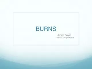
BURNS - MANAGEMENT. LOVYA GEORGE 2002 BATCH. PRE-HOSPITAL CARE. Remove from the source of injury Ensure rescuer safety Cool the burn wound, avoid hypothermia All rings, jewelry,watches and belts should be removed. EMERGENCY CARE. Airway Breathing Circulation Disability Exposure.

E N D
BURNS - MANAGEMENT LOVYA GEORGE 2002 BATCH
PRE-HOSPITAL CARE • Remove from the source of injury • Ensure rescuer safety • Cool the burn wound, avoid hypothermia • All rings, jewelry,watches and belts should be removed
EMERGENCY CARE • Airway • Breathing • Circulation • Disability • Exposure
AIRWAY • Burned airway creates symptoms by swelling • Early intubation in suspected airway burn is safest • Clues of airway burn- blisters on hard palate, burned nasal mucosa, loss of hair in the nose and deep burns around the mouth and neck.
BREATHING • inhalational injury-due to smoke inhalation • Suspect in those trapped in fire, presence of soot in nose • Symptoms develop as late as 24 hrs to 5 days
Inhalational injury • Physiotherapy • Nebulisers • Warm humidified O2 • Blood gas measurements • IPPV if condition deteriorates • COHb of > 10% high inspired O2
Metabolic poisoning Suspect in those trapped in fire. • Mechanical block to breathing. Full thickness burns on chest wall. Do escharotomy.
OUTCOME • Percentage of surface area involved • Depth of burns • Presence of an inhalational injury • Age • Associated medical problem or other injuries
ASSESSMENT OF SIZE • In small burns patients hand can act as a guide.The area of hand is roughly 1% of TBSA. • Wallace’s rule of nine • LUND BROWDER CHART
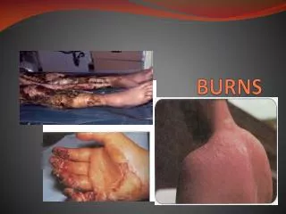
Assessment of Depth • History- temperature, time and burning material. • Superficial burns have capillary filling. • Deep partial thickness burn do not blanch,and has some sensation. • FTB is hard and leathery and has no sensation.
FLUID THERAPY • First fluid resuscitation formula by Evans • The principle is to maintain Intravascular vol. at a state sufficient to perfuse not only all vital organs but also the peripheral tissues. • Fluid loss depends on BSA involved.
Cont… • IV fluid resuscitation appropriate : • CHILD > I0% TBSA ADULT >15% TBSA FLUIDS Ringer lactate human albumin FFP Hypertonic saline Max. fluid loss – first 8 hours
IV ACCESS • Upper limb preferred • LL more chance of septic thrombosis • <50%- peripheral venous cannula • >50% -central venous line
FORMULAS • Parkland • Evans • Staler • Brooke • Modified Brooke • Metrohealth
PARKLAND FORMULA • Dr.Charles Baxter of PARKLAND Hospital • Total vol. in first 24 hrs= 4 ml/kgBody Weight/% of burn TBSA • Half in first 8hrs • Next half in next 16 hrs
TIME DEPENDENT VARIABLES SHOULD BE CALCULATED FROM TIME OF BURNS • For next 24 hrs • 30-60% of plasma loss • CHILDREN – maintenance fluid DS • up to 10 kg-100ml/kg/24 hrs • 50ml/kg for next 10 kg • more than 20kg-20ml/kg/24 hrs
MONITORING • URINE OUTPUT • b/w 0.5-1 ml/Kg/Hr • if below this-increase infusion by 50% • below this+signs of hypoperfusion -10ml/Kg Bolus • Decrease if >2ml/kg/hr
Role of COLLOIDS • Usually in 2nd 24 hr • After capillary leak has subsided • Mostly in • Burns > 40% • heart diseases • inhalational injuries • geriatric age group
Muir and Barclay Formula • fluid -during the first 36 hours after a major burn… • 0.5ml/Kg body weight/5 TBSA • Each infusion volume is given as follows: • first 12 hours - 3 infusions at 4 hour intervals • second 12 hours - 2 infusions at 6 hour intervals • third 12 hours - 1 infusion • ALBUMIN is the resuscitation fluid.
BROOKE FORMULA • FIRST 24 HOURS • RL 1.5ml/kg/%burn • COLLOID – 0.5ml/kg/% • GLUCOSE IN WATER – 2000ml • SECOND 24 HOURS • RL - Half to three fourth of first 24 hour requirement • COLLOIDS – • 2000 ml
EVANS FORMULA FIRST 24 HOURS N.S. – 1ml/kg/%burns COLLOID – 1ml/kg/%burns GLUCOSE IN WATER – 2000ml SECOND 24 HOURS HALF OF FIRST REQUIREMENT
SLATER FORMULA • RL – 2L/24 HOUR • FFP – 75ml/kg/24hour MONAFO FORMULA • Hypertonic saline • VOLUME – to maintain urine output at 30ml/L
DEMLING FORMULA • FFP & DEXTRAN • Dextran 40 in saline - 2ml/kg/h • RL – To maintain output at 30ml/hr • FFP – 0.5ml/kg/h for 18hrs
Fluid reqd will be more than indicated in • Electric burns • Burned while drunk • Delayed resuscitation • Gross soft tissue involvement
Volume sensitive pts will be • Those above 50 yrs • Less than 2 yrs • With cardio-pulmonary disease
Tetanus prophylaxis • Escharotomy- in circumferential FTB
ESCHAROTOMY • Are releases of burn eschar performed at bedside with a scalpel or electrocautery unit. • Need arises in second and third degree burns. • wounds encompassing the circumference of an extremity interfering with peripheral circulation to the limb by a tourniquet effect.
Oedema beneath eschar impedes venous outflow and affects arterial inflow to limbs. • Truncal eschar decrease ventilation by limiting chest expansion. • In the neck oedema may obstruct trachea
Indications • Clinicalsigns • absence of pulse • pain in the limb • cyanosis,impaired capillary refilling • Doppler signals indicating decreased or absent flow • Compartment pressure > 40mm Hg • Oxygen saturation <95%
AIM • To release pressure over involved deeper tissues and restore circulation. • TECHNIQUE • Ward procedure • No need of anaesthesia
Escharotomies • In a circumferentially burned limb - along the mid lateral or mid medial line. • should extend from proximal to distal margin of burn area. • should extend through the entire depth of eschar.
IN chest • bilaterally in the anterior axillary line extending from clavicles to coastal margins. • if chest expansion not adequate join with transverse incision along costal margin.
Bleeding must be controlled • Elevation of limb to prevent oedema • Complications • blood loss • release of anaerobic metabolites causing transient hypotension • If escharotomy does not restore blood flow fasciotomy is required
Wound dressing • Superficial PTB heal irrespective of the dressing • Borderline deep dermal burns may heal without scar if properly dressed with suitable dressing
Topical antimicrobial dressing • Simplest method of treating superficial wound is by exposure • Permeable wound dressing eg: fixamol or Mefix. • Vaseline impregnated gauze or a fenestrated silicone sheet.
Hydrocolloid dressings especially useful in mixed depth burns. • Moist environment good for epithelialisation. • Eg: Duoderm
Biological dressings Synthetic and natural • Biobrane,Trancyte,integra, amniotic membrane, allograft xenograft etc. • Good healing environment • No need to change • Useful in superficial burns.
Biobrane • Consist of collagen coated silicone manufactured into a sheet • Provides a barrier to moistureloss,does not require dressing changes • Epithelium is complete under biobrane sheet it is easily peeled off the wound
TRANSCYTE • Similar to biobrane with addition of growth factors from lysed fibroblast grown in culture • INTEGRA • A product that combines a collagen matrix (dermal sustitute) with silicone sheath outside layer (epidermal substitute) • BIOLOGICAL DRESSINGS • xenografts from swine • allografts from cadaver donor
FTB and obvious deep dermal burn – require operative treatment for excision and skin grafting • Early excision is preferred rather than wait for eschar to separate.
advantages Decrease • Hospital stay • Need for painful debridements • Infectious complications • Hypermetabolism • Scarring
Till then managed by antibacterial dressing to prevent bacterial colonisation • Agents : earlier staphylococci and streptococci • Now pseudomonas
Topical treatment • 1% Silver sulphadiazene • Method : exposure or single layer dressing • Broad coverage, also to pseudomonas and MRSA. • Painless,easy • Limited eschar penetration
.5% Silver nitrate solution • Occlusive dressing • Change in 2 – 4 hrs • Black staining • Mafenide acetate 5% • Exposure • Painful
Serum nitrate • Bacitracin • Neomycin, • Polymyxin B • Mupirocin etc
Escharectomy • Excision of whole or part of an eschar • Burn eschar is a necrotic tissue • Can lead on to systemic infections and intoxication
Early excision and grafting is done in full thickness and deep dermal burns.(except <4 cm square). • Can be done in first week when the patient is heamodynamically stable. • Can be done serially or in a single operation