BURNS
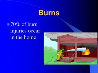
BURNS. BURNS . Burn injury and the number of deaths - dropped in the past 10 years -decrease is from: -use of smoke detectors -creation of regional burn centers -national focus on safety -occupational safety mandates. Causes.

BURNS
E N D
Presentation Transcript
BURNS • Burn injury and the number of deaths • - dropped in the past 10 years • -decrease is from: • -use of smoke detectors • -creation of regional burn centers • -national focus on safety • -occupational safety mandates
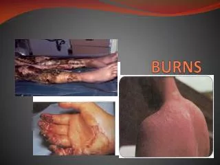
Causes • -Thermal or nonthermal causes. • -Thermal burns • -most common type of burn injury • -caused from heat • -flames, scalds, thermal energy • -Nonthermal burns • -electricity, chemicals, and radiation.
Causes • Skin destruction • -depends on the burning agent • -condition of the skin before injury • -duration of the person’s contact with the agent
Severity of burns • The factors that determine the severity of a burn are: -Percentage of the body surface area burned. - Age -Specific location of the burn. -Cause -Other diseases -Depth of the burn. -Injuries
Depth of burns/Classification • Superficial thickness injuries • Partial-thickness injuries • Full-thickness injuries • - graphically describe the burn • -depth and severity of the tissue injury See AHN p. 95, Table 3-3 for descriptions of the burn classifications. -If you use only the visual characteristics of the burn wound, it would not provide an accurate assessment of how much damage might have been caused.
Percentage Estimates • The RULE OF NINES • - determines the total body surface area (BSA) burned. See p. 106, FIGURE 3-22. • The “rule of nines” divides the body into multiples of nine. -Head to neck 18% -Arm (shoulder to fingertips) 9% each -Anterior trunk 18% -Posterior trunk 18% -Leg (groin to toe) 14% each
Age considerations • Percentage of body area burned in infants and children -the surface area of the child’s head is larger • Increased risk to develop circulatory • -adults with cardiac disease • -the very old • -the very young overload.
Burns • Dramatic changes • -first few minutes to the first 12-24 hours
Extent of the burns? • -greater the 20% • -cause massive evaporative of water • -fluid losses into the interstitial space • -capillaries dilate (hypermeablitiy) for 24 hours • -fluid shifts from the capillaries to the • interstitial spaces • -causes edema and blistering (third spacing) • -cells become dehydrated • -hypovolemic shock starts • - hypotension and decreased renal flow
Three stages of medical treatment • 1. Emergent Phase (Stage 1) • Decreased volume and shock • -occur up to 48 hours after being burned.
Three stages of medical treatment • 2. Acute Phase (Stage 2) • -48-72 hours after a burn • -circulatory overload • - secondary to fluid shifting back from the interstitial • spaces to the capillaries • - increased urine output • -“diuretic stage”
3. Rehabilitation Phase (Stage 3) • -wound treatment begins • -slowly returns to as normal status as • possible
Complications • Carbon Monoxide (CO) poisoning • -Person in an enclosed area during a fire • -CO displaces O2 from hemoglobin • -Don’t’ rely on oximeters • - can’t distinguish from oxyhemoglobin and • carboxyhemoglobin • -Early signs- • -headache • -nausea • -vomiting • - unsteady gait • -Treatment- 100% oxygen
Smoke Inhalation • Inhaling chemicals produced by the fire • Damages- • -celia and mucous membranes of the respiratory • tract • Symptoms- • -several hours after the initial burn • High risk for patients • -upper chest, neck and face burns
Smoke Inhalation • -hoarse voice -gutteral breath sounds • -productive cough -redness/swelling • -sooty sputum -nasal or oral pharynx • -singed nasal hairs • -agitation • -tachypnea • -flaring nostrils • -intercostal retractions • - grunting
Smoke Inhalation • Treatment • -establish airway • -initiate oxygen • -may need intubation
Shock • Emergent phase • -fluid shifting from the capillaries to the interstitial • spaces. • Requires fluid resuscitation (IV fluids) • -Adults-greater then 20% of their body surface • -Children-10 % • -Older then 55 • -Younger then 14 years • -Cardiac, pulmonary disease or diabetic • -Electric burns
IV fluid therapy • -central line of Lactate Ringers • -amount of fluid given • -body weight • -percentage of body surface burned. • Foley catheter • -monitors urine output. • -30-50 cc/hour urine output • -maintain adequate renal function • Airway • -continue to maintain • -vital signs monitored
Infection • Most common cause of death in the first 72 hours in burn victims • Nursing implications • -erythema, odor, green or yellow exudate • -wound culture and sensitivity • -topical bacteriostatics -capillaries are coagulated by the burns
Protective Isolation • -gown, mask, cap and glove • -dressing changes require strict surgical aseptic • techniques.
Immediate Medical Management • 1. Establish an airway • -Oxygen -intubated to ensure a patent airway • 2. Initiate fluid therapy • -Insert a central IV line • -Ringers Lactate IV immediately • -the amount depends on: • -body weight and the • -percentage of the body surface area burned
3. Renal function and urine output -insert a foley catheter -maintain a 30-50cc/hour urine output to perfuse the kidneys -adjust the IV fluid to maintain adequate urine output • 4. Pain control -Morphine IV -small doses given frequently
-3-5mg IV every 5-10 minutes until pain relived • -Children- 0.1-0.2 mg/kg every 2-4 hours PRN • -Hypovolemic -effects of analgesic may increase • -Monitor for respiratory depression • -Fentanyl may be an alternative if the client is • allergic to Morphine
6. Body temperature -chilling -secondary to the skin being left open to the air for wound healing. -keep room at 85 degrees and humidity at 40-50% -light and heat lamps (use caution) • 7. Infection control -Tetanus immunization if client is not up to date, -Wound infections-topical bacteriostatics -Systematic infections (pneumonia) -IV antibiotics.
Recovery Phase • - 10 days to several months depending on severity of the burns • -72 hours after a burn injury • -increased metabolism • -decreased urine output • -decreased edema • -Goals • -treat burn wounds • -prevent and manage complications
Prevent Complications • -Infections • -heart failure • -renal failure • -extremity contractures • -paralytic ileus • -Curling’s ulcer
Wound Debridement • Debridement • -removes the damaged tissue/debris from a • wound or burned tissue • -prevents infection • -promotes healing • Partial thickness wounds • -debrided twice a day • -topical antibiotic • -dressing applied
Eschar removal • Black leathery crust -forms over burned tissue -holds in micro-organisms -causes infection • Escharectomy- -cutting down to the healthy tissue -chest expansion is restricted -burns around the chest, arms or legs
Debridement • -Helps with regeneration of the tissues • -Enzymes • -applied topically • -chemically debride the eschar • -Hydrotherapy • -softens the eschar with water • -makes debridement less painful • -promotes range of motion the extremities • - preventing contractures
Debridement • Failure to debride • -increased the chance for infection • -delays healing • -increases scarring
WOUND CARE • -Severity of the burn • -Open (exposure) method -burns of the face, neck, ears, and perineum -cleaned and exposed to air -hard crust forms -regeneration of tissue occurs -advantages : -wound can be observed -body part is not restricted -circulation is not compromised -exercises can be performed more easily
Pain Control • Changing the dressing will be PAINFUL!!!!! • -Analgesics-given at least 30 minutes before dressing changes • -IV Morphine • -Remove dressings after hydrotherapy
Rehabilitation • -Less the 20% BSA remains burned -Physical and Occupational Therapy work -improve endurance, strengthening and independence in ADLs • Nursing Implications • -realistic short term goals-keep the client motivated -encourage to verbalize feelings about his changed body image
Surgical Options • Skin Grafts- • -Prevents the scar tissue • -disfigurement • -and loss of mobility • -Required for burns • -disrupted the epidermis • -most of the dermis
Surgical Options • -Promotes healing • -Prevents infection • -First 3 weeks after a burn • -4 types of grafts -auto graft -homograft -heterograft -synthetic graft
Auto graft • Surgical transplantation of tissue from one part of the body from the same person
Homograft • Surgical transfer of tissue from two genetically different individuals of the same species • -a temporary graft can be from a cadaver
Heterograft • Tissue from another species • -Temporary graft
Synthetic Graft Made from a variety of materials such as neonatal human fibroblast cells TransCyte developed in 1997 -applied only once -temporary covering -protect against fluid loss -decreases the chance of infection
Methods of application of grafts • Pedicule method -partially attached to the donor site and the other portion is attached to the burn site • Free standing method -tissue is completely removed from the donor site and attached to the burn site
Client education • 1. Do not to remove the dressing until the physician orders the removal. • 2. Report bruising or fluid collection under the graft to the physician. • 3. Protect the skin graft from sunlight/use sunscreen to the graft site for 6 months after the surgery. • 4. Use lotion to the skin graft site for 6-12 months. • 5. Wear elastic stocking when having skin grafts to the lower extremities for 4-6 months
Pharmacology Anti-Infectives, Antiseptics and Germicides- • -Topical medications -prevent wound infections • Types- • Sulfamylon Silvadene • Silver Nitrate Furacin • Gentamycin Neomycin