Download
1 / 74
750 likes | 992 Views
BURNS Early management issues. Epidemiology. Approx 135 000 total burn injuries in Oz in 2001. 2% of all injury hospitalisations 6000 children to A&E with burns each year 20-25 children die each year from burns 13 000 hosp. in NSW between 95 and 99, 40% children.

E N D
Epidemiology • Approx 135 000 total burn injuries in Oz in 2001. • 2% of all injury hospitalisations • 6000 children to A&E with burns each year • 20-25 children die each year from burns • 13 000 hosp. in NSW between 95 and 99, 40% children. • Declining trend of burn-related deaths in NSW from 90s to early 2000s. • Gender 60% men • Age – death higher among the elderly; late teens to mid 40s most commonly affected.
Types of burns • Thermal • Chemical • Radiation • Electrical
Thermal burns • Fire (46%) • Flash, flame • Scalding (32%) • Liquids, grease, stream • Contact (8%) • (Electrical – voltage > 1000 V) (4%)
Simple applied physics • Temperature (energy) • Duration / exposure time • Medium • Skin thickness (age) (intrinsic structure of tissue) • Heat dissipation (blood flow)
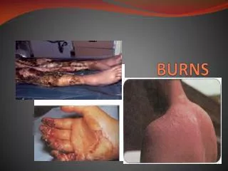
Depth of burn injury • Classified in degrees of injury based on the amount of epidermis and dermis injured. At present, depth is estimated by physical appearance, pain, and skin texture or pliability. • First-degree burninvolves only the thin outer epidermis and is characterized by erythema and mild discomfort, healing rapidly. • Second-degree burnsare defined as those in which the entire epidermis and variable portions of the dermis are destroyed. Subdivided into superficial second-degree burn and deep dermal(or deep second-degree) burn • Full-thickness (or third-degree) burnoccurs with destruction of the entire epidermis and dermis, leaving no residual epidermal cells to repopulate the burned area. The portion of the wound not closed by wound contraction will require skin grafting.
Prognosis • Overall survival rates from all burns 95% • Greatly improved survival over last 50 years • Shock Sepsis Inhalation / pneumonia • Pneumonia now greatest cause of mortality • % TBSA burnt + age = mortality • Multidisciplinary approach and specialised burns centres
Indicators of poor prognosis • Extremes of age • %TBSA • Severe inhalation injury and ventilator dependency • Combination of inhalation injury with cutaneous burn • Co-existent trauma • Acute renal injury / elevated creatinine • Poor pre-morbid health status • Sepsis / pneumonia (mortality increased with 40%) • Thrombocytopenia (< 20 000) • Elevated serum lactate and / or base deficit.
NSW Referral centres • Concorde • Royal North Shore • Westmead (under 16 years of age)
Who to refer? • Partial/full thickness burns in adults >10% TBSA. • Partial/full thickness burn in children > 5% TBSA. • Burns to the face, hands, feet, genitalia, perineum and major joints. • Chemical burns • Electrical burns • Burns with concomitant trauma • Burns in patients with pre-existing medical conditions that could adversely affect patient care and outcome • Children with suspected non-accidental injury • Pregnancy with cutaneous burns
Who to retrieve? • Any intubated patients • Head and neck burns • Partial and full thickness burns > 10% in children / > 20% in adults • Burns with significant co-morbidities • Associated trauma • Significant pre-existing medical disorder • Electrical conduction injury with cutaneous burns • Chemical injury with cutaneous burns
Management • Trauma team • Handover from paramedics • Details important – when, where, how, what etc. • First aid / treatment this far • Vital signs
Trauma call A burn patient is a trauma patient; therefore, other injuries should be expected and sought
Dramatic physiologic and metabolic changes over the course of the injury state. • Three phases of burns: 1) Resuscitation phase (0 to 36 hours) characterised by cardiopulmonary instability 2) Post resuscitation phase (2 to 6 days) 3) Inflammation / infection phase (7 days to wound closure)
Airways & Breathing Pathophysiology of early changes 1) Inhalation injury complex • Toxic compounds absorbed • Upper airway obstruction • Chemical irritation / injury to airways and lung parenchyma 2) Burn injury (external) to face and neck 3) Burn injury (external) involving the thorax
Smoke inhalation injury complex • Pulmonary insufficiency caused by the inhalation of heat and smoke is the major cause of mortality in the fire-injured person, accounting for more than 50% of fire-related deaths. Acute upper airway obstruction occurs in 20-33% of hospitalised burn patients with inhal. injury. • Many new synthetics in home furnishings and clothing have resulted in a much more complex form of injury, due to the extremely toxic combustion products of these advances in technology. • A closed space fire can result in a severe hypoxic insult as well as lung damage from the inhalation of toxic fumes. • The exposure time, the concentration of fumes, the elements release and the degree of concomitant body burn are critical variables. • These factors cause a very complex injury with morbidity and mortality risks, especially when combined with a body burn.
Carbon monoxide toxicity • One of the leading causes of death in fires. • Basic by-product of (incomplete) combustion. • Rapidly transported across the alveolar membrane. Preferentially binds with the haemoglobin molecule in place of oxygen (*200). Shifts the Hb-oxygen curve to the left, thereby impairing oxygen unloading at tissue level. • Tissue hypoxia. Also binds to myoglobin. Can also saturate the cell, bind to cytochromeoxidase and thereby impair mitochondrial function and ATP production.
Carbon Monoxide Toxicity How to diagnose? • ABG • High COHb • Unexplained metabolic acidosis • Low SpO2 for PO2
Cyanide toxicity • Cyanide toxicity presents in a very similar fashion to carbon monoxide, with severe metabolic acidosis and obtundation in severe cases. • Normal levels < 0.1 mg/L • Binds to cytochrome c oxidase and disrupts the electron transport chain, inhibiting aerobic metabolism and depleting cells from ATP. • Diagnosis is more difficult because cyanide levels are not always readily available or very reliable.
Treatment CO toxicity • Oxygen and supportive care. • Hyperbaric oxygen • T1/2 room air – 90 minutes. T1/2 FiO2 1.00 – 30 minutes. Treatment cyanide toxicity • Cardiopulmonary support is usually sufficient treatment, since the liver via the enzyme rhodenase will clear the cyanide from the circulation. • Sodium nitrite is used (300mg intravenously over 5 to 10 minutes) in severe cases (confirm levels). • Hydroxycobalmin and thiosulphate.
Upper airway obstruction from tissue oedema • Direct heat injury caused by the inhalation of air heated to a temperature of 150O C or higher ordinarily results in burns to the face, oropharynx, and upper airway (above the vocal cords). • Heat immediate injury to the airway mucosa with oedema, erythema, and ulceration. Anatomically these changes may be present shortly after the burn, but clinical signs may not occur till 12-18 hours after injury. • Inhalational injury + body burn much higher risk of oedematous airway obstruction due to fluid resuscitation given and the release of inflammatory mediators from the burned skin. • Burn to face or neck marked anatomic distortion and, in the case of the deep neck burn, external compression on the larynx. Third degree burn of the neck is particularly bad Minimal external oedema due to the non-elastic burn No external expansion. Massive intraoral / pharyngeal oedema • Increased secretions • Oedema resolves around day 4-5 unless there is extensive and deep injuries.
Symptoms & signs of obstruction • Upper airway noise (turbulent airflow), dyspnoea, increased work of breathing, anxiety, stridor and eventually cyanosis. • Difficult to distinguish noise from a narrowed airway from that caused by increased oral and nasal secretions due to smoke irritation. • The airway oedema and the external burn oedema process have a parallel time course so that by the time symptoms of airway oedema develop, external and internal anatomic distortion will be extensive.
How to confirm airway involvement if in doubt? How to determine degree of involvement? • Signs of facial burn / erythema, swollen lips, singed facial hair, carbonaceous sputum. • Serialfibreopticbronchoscopies/ laryngoscopies. • Remember oedema is progressive up until 18 hours post injury.
Treatment • Intubate early if indicated • Otherwise close monitoring and regular reviews are essential while... • Positioning the patient to minimise head/neck swelling • Careful not to overhydrate and promote oedema • Analgesia • Escharotomy (patient usually intubated by this stage) • More to follow...
CHEMICAL BURN TO UPPER AND LOWER AIRWAYS • Generally much more serious than that produced by heat alone. • Exposure to toxic gases contained in smoke PLUS carbon particles coated with irritating aldehydes and organic acids • Injury to both upper and lower airways. • The location of injury will depend on the duration of exposure, the size of the particles, and the solubility of the gases.
The unconscious patient loses airway protective mechanisms, resulting in a more severe injury to the lower airways when continuing to inspire. • Water-soluble gases such as ammonia, sulphur dioxide and chlorine react with water in the mucous membranes to produce strong acids and alkalies irritation, bronchospasm, mucous membrane ulceration and oedema. Severe impairment of the ciliary mechanism impaired removal of particles and mucus. • Lipid-soluble compounds, e.g. nitrous oxide, phosgene, hydrogen chloride, and various toxic aldehydes, are transported to the lower airways on carbon particles that, in turn, adhere to the mucosa. All these agents produce cell membrane damage. • Alveolar oedema is not a major component of the early disease state.
Symptoms may be absent on admission. The magnitude of the degree of injury evident after 24 to 48 hours. • Early symptoms usually consist of bronchospasm manifested as wheezing and bronchorrhoea. Coughing. Sometimes confused with pulmonary oedema. • Marked decrease in lung compliance and increased work of breathing. Impaired clearance of secretions. • V/Q mismatch with increased A-a gradient. • Injury at the alveolar level is usually fatal.
Diagnosis • History – exposure, confined space? • Symptoms & signs • High HbCO • Laryngoscopy • Absence of upper airways injury (serial reviews) usually means absence of lower airway injury. • Bronchoscopy (if intubated) • Xenon scan (not in acute settings)
Treatment • Aggressive approach to upper airway maintenance and pulmonary support, which includes maintenance of small airways patency and removal of soot and the mucopurulent secretions. • I.e. very likely to need intubation. • PEEP to maintain small airway patency and an adequate FRC. Prevention easier than treating. • Early intubation and PEEP have been reported to decrease pulmonary deaths after severe burns and smoke inhalation. • Tube size – minimal 7 mm for adults. • Humidified oxygen • Elevation of the patient’s head and chest 20 to 300 is also helpful. • Careful well-monitored fluid resuscitation • Bronchodilators for bronchospasms. • Anticholinergics to minimize bronchorrhoea + bronchodilator effect? • No role for AB and steroids.
IMPAIRED CHEST WALL COMPLIANCE • Respiratory excursion can be markedly impaired by a burn to the chest wall. Most evident with a circumferential third degree burn with loss of elasticity in the chest wall due to the burn tissue . Increased WOB to maintain functional residual capacity and an adequate tidal volume. • Oedema from a second degree burn is also sufficient to alter lung mechanics (axillae and lateral chest walls). • Compressed intrathoracic volume significant V/Q mismatch, atelectasis, and hypoventilation. Maximum respiratory effort is required just to maintain adequate gas exchange.
Symptoms may not be clearly evident until oedema formation peaks at about 10 to 12 hours. • In the combined chest burn and inhalation injury it is very difficult to distinguish the degree of impairment in total lung compliance due to the increased airway oedema and bronchospasm compared with that due to the impaired chest wall. Treatment • Positioning and judicious fluid resuscitation. • NIV or mechanical ventilation. • Escharotomy (early if circumferential 3rd degree).
A&B Summary of early management • High flow 70-100% oxygen to all patients • Assess airway and surrounding tissues • Intubate (RSI) if indicated • ? In-line immobilisation of neck • Risk factors for (early) intubation: • Unconsciousness at scene • Fire in confined space • Facial burns singed facial hair, soot in nostrils or sputum, facial erythema. • Voice changes or “lump in throat” • Elevated carbon monoxide levels on ABG or respiratory failure. • Assess breathing and thorax • Intervention? • Continue primary survey and obtain monitoring and ABG results. CXR
Secondary survey • If not intubated yet • Other injuries identified? • Time, equipment and appropriate staff for laryngoscopy? • Positioning of patient • Assist with clearance of secretions • Fluid management • Chest wall excursion • ?Role of NIV • Bronchodilators if wheezing
Criteria for intubation (NSW Health) • Clinical evidence of possible airway compromise: • Head and neck burns/scalds with increased swelling • Stridor, hoarse voice, swollen lips • Carbonaceous material around or in the mouth, nose or sputum • Singed facial, head or nasal hairs. • Intubate early • If patient unconscious • If there are head and neck burns with obvious swelling • If the patient is to be transported and meets any of the above criteria. • If there are other clinical symptoms and signs and ABG results are indicative of respiratory dysfunction.
Potentially five major pulmonary problems: • Continued Upper Airway Obstruction • Decreased Chest Wall Compliance • Tracheobronchitis from Inhalation Injury • Pulmonary Oedema • Surgery - and Anaesthesia-Induced Lung Dysfunction 30-70% of patients with inhalational injury will develop ventilator-associated pneumonia.
Continued upper airway obstruction Pathophysiology • Continued airways oedema • Mucosal damage with slough • Increased oral secretions • Bacterial colonization Treatment • Keep intubated until oedema resolves • Head elevated position • Avoid excessive tube motion • Vigorous oral hygiene (+/- Nystatin if on antibiotics) • Avoid cuff over-inflation • Consider tracheostomy When can the patient be extubated?
Decreased chest wall compliance • Not completely eliminated by escharotomy • Continuous swelling for days • High PEEP can affect haemodynamics • More difficult to manage during GA Treatment • Continue supportive care and mechanical ventilation. • Care with fluid adm. • Early surgical management of full thickness burns
Tracheobronchitis Pathophysiology • Ongoing mucosal injury (degree and duration depending on chemical exposure) • Increased secretions / bronchorrhoea and impaired ciliary function • Bronchospasm • Interstitial oedema • Necrosis and slough • Airway plugging, atelectasis and hypoxaemia • Increased risk of infection (colonisation inevitable) • Tracheobronchitis bronchopneumonia
Clinical findings • Sputum changing from loose to purulent • Evidence of necrotic tissue in sputum • Wheezing, ronchi, creps +/- bronchial breathing • Increased work of breathing • Altered gas exchange • Bronchoscopic findings • Infiltrates on radiographs: Late finding Treatment • Aggressive pulmonary toilet with frequent postural drainage (consider rotation bed) ; physiotherapy. • Infection surveillance (daily sputum/ETT samples) • Antibiotics when indicated (not prophylactic ) • Inhaled bronchodilators • Inhaled N-acetylcysteine? • Positive pressure to maintain FRC • Aggressive diuresis to correct airways oedema not shown to work