Download
1 / 34
400 likes | 668 Views
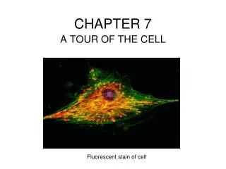
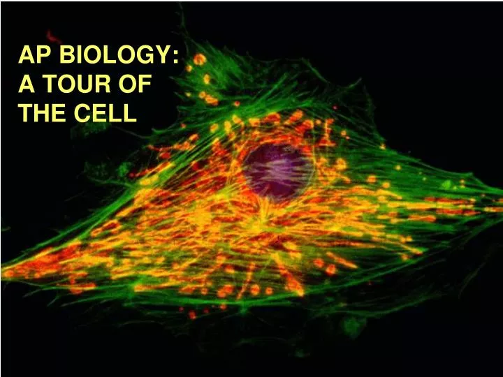
AP BIOLOGY: A TOUR OF THE CELL. ANIMAL CELL. PLANT CELL. Nucleus. Contains most of the genes in the eukaryotic cell Enclosed by a nuclear envelope —a double membrane (lipid bilayer) perforated by pores which regulates the entry and exit of certain large particles

E N D
Nucleus • Contains most of the genes in the eukaryotic cell • Enclosed by a nuclear envelope—a double membrane (lipid bilayer) perforated by pores which regulates the entry and exit of certain large particles • Envelope is lined by a nuclear lamina, a net-like array of protein filaments that maintain the shape of the nucleus
Nucleus Cont. • Within the nucleus, DNA is organized with proteins into chromatin. This chromatin coils up into chromosomes during reproduction • The nucleolus a dark area of the nucleus, is in charge of the synthesis and assembly of rRNA (ribosomal RNA) to be imported into the cytoplasm and become subunits of ribosomes • The nucleus directs protein synthesis by making mRNA and sending it into the cytoplasm
Ribosomes • Made of rRNA and proteins • Carry out protein synthesis • Made of 2 subunits (large subunit & small subunit) • Found in large numbers in cells with high amounts of protein synthesis • Build proteins in 2 cytoplasmic locales: • Free Ribosomes: are suspended in the cytosol; makes proteins that are used in the cytosol • Bound Ribosomes: are attached to the endoplasmic reticulum or the nuclear envelope; makes proteins that are used in the endomembrane system or for export • Cells can adjust the number and type of ribosomes depending on metabolism changes
Endomembrane System • Group on internal membranes related to each other through direct contact or by transfer of membrane segments, vesicles • Includes: • Nuclear envelope • Endoplasmic Retiuclum • Golgi apparatus • Lysosomes • Various vacuoles • Plasma membrane (not actually endomembrane but still connected)
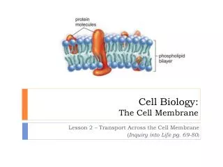
Endoplasmic Reticulum • Membranous labyrinth made up of more than half the total membrane in most eukaryotic cells • Made of membranous tubules and sacs called cisternae • The ER membrane is continuous with the nuclear envelope and the cisternal space of the ER is continuous with the space between the two membranes of the nuclear envelope. • There are two regions of ER that differ in structure and function. • Smooth ER looks smooth because it lacks ribosomes. • Rough ER looks rough because ribosomes (bound ribosomes) are attached to the outside, including the outside of the nuclear envelope.
Smooth ER • Enzymes of smooth ER synthesize lipids, including oils, phospholipids, and steroids. • The smooth ER also catalyzes a key step in the mobilization of glucose from stored glycogen in the liver. • Other enzymes in the smooth ER of the liver help detoxify drugs and poisons.
Rough ER • Rough ER is abundant in cells that secrete proteins. • As a polypeptide is synthesized by the ribosome, it is threaded into the cisternal space through a pore in the ER membrane. • Many of these polypeptides are glycoproteins • These secretory proteins are packaged in transport vesicles • Rough ER is also a membrane factory. • Membrane bound proteins are synthesized directly into the membrane. • Enzymes in the rough ER also synthesize phospholipids from precursors in the cytosol. • As the ER membrane expands, parts can be transferred as transport vesicles to other components of the endomembrane system.
Golgi Apparatus • The Golgi apparatus consists of flattened membranous sacs - cisternae - looking like a stack of pita bread. • finishes, sorts, and ships cell products • center of manufacturing, warehousing, sorting, and shipping. Tags, sorts, and packages materials into transport vesicles. • Many transport vesicles from the ER travel to the Golgi apparatus for modification of their contents. • Found most in cells specialized for secretion. • One side of the Golgi, the cis side, receives material by fusing with vesicles, while the other side, the trans side, buds off vesicles that travel to other sites.
Lysosomes • The lysosome is a membrane-bounded sac of hydrolytic enzymes that digests macromolecules. • Lysosomal enzymes can hydrolyze proteins, fats, polysaccharides, and nucleic acids. • While rupturing one or a few lysosomes has little impact on a cell, massive leakage from lysosomes can destroy an cell by autodigestion, known as apoptosis • Lysosomes can fuse with food vacuoles, formed when a food item is brought into the cell by phagocytosis. • Lysosomes can also fuse with another organelle or part of the cytosol, autophagy, renews the cell. • Several inherited diseases affect lysosomal metabolism. • Pompe’s disease in the liver and Tay-Sachs disease in the brain.
Vacuoles • Vesicles and vacuoles (larger versions) are membrane-bound sacs with varied functions. • Food vacuoles, from phagocytosis, fuse with lysosomes. • Contractile vacuoles, found in freshwater protists, pump excess water out of the cell. • Central vacuoles are found in many mature plant cells. • The functions of the central vacuole include stockpiling proteins or inorganic ions, depositing metabolic byproducts, storing pigments, and storing defensive compounds against herbivores. • The membrane surrounding the central vacuole, the tonoplast, is selective in its transport of solutes into the central vacuole.
Other Membrane Bound Organelle • Mitochondria and chloroplasts are the energy transformers of cells. They convert energy to forms that cells can use for work. • Mitochondria are the sites of cellular respiration, generating ATP from the catabolism of sugars, fats, and other fuels in the presence of oxygen. • Chloroplasts, found in plants and eukaryotic algae, are the sites of photosynthesis—convert solar energy to chemical energy and synthesize new organic compounds from CO2 and H2O. • Mitochondria and chloroplasts are NOT part of the endomembrane system. • Their proteins come from free ribosomes and from their own ribosomes. • Both have small amounts of DNA that direct the synthesis of their own polypeptides • Mitochondria and chloroplasts grow and reproduce as semiautonomous organelles.
Mitochondria • Mitochondria have a smooth outer membrane and a highly folded inner membrane, the cristae. • This creates a fluid-filled intermembrane space between them. • The cristae present ample surface area for the enzymes that synthesize ATP. • The inner membrane encloses the mitochondrial matrix, a fluid-filled space with DNA, ribosomes, and enzymes. • They are found in almost ALL eukaryotic cells • Their number correlates with the aerobic activity of the cell
Chloroplasts • The chloroplast produces sugar via photosynthesis. • Chloroplasts gain their color from high levels of the green pigment chlorophyll. • Chloroplasts are found in leaves and other green structures of plants and in eukaryotic algae. • Inside the innermost membrane is a fluid-filled space, the stroma, in which float membranous sacs, the thylakoids. • The stroma contains DNA, ribosomes, and enzymes for part of photosynthesis. • The thylakoids, flattened sacs, are stacked into grana and are the membranes used for converting light to chemical energy.
Other Plastids • The chloroplast is one of several members of a generalized class of plant structures called plastids. • Amyloplasts store starch in roots and tubers. • Chromoplasts store pigments for fruits and flowers
Peroxisomes • Peroxisomes generate and degrade H2O2 in performing various metabolic functions • contain enzymes that transfer hydrogen from various substrates to oxygen • An intermediate product of this process is hydrogen peroxide (H2O2), a poison, but the peroxisome has another enzyme (catalase!) that converts H2O2 to water and oxygen. • Some peroxisomes break fatty acids down to smaller molecules that are transported to mitochondria for fuel. • Others detoxify alcohol and other harmful compounds. • They form not from the endomembrane system, but by incorporation of proteins and lipids from the cytosol.
Cytoskeleton • The cytoskeleton is a network of fibers extending throughout the cytoplasm. • The cytoskeleton organizes the structures and activities of the cell. • providing structural support to the cell, the cytoskeleton also functions in cell motility and regulation • provides mechanical support and maintains shape of the cell. • provides anchorage for many organelles and cytosolic enzymes. • plays a major role in cell motility. • dynamic, dismantling and reassembling to change cell shape. • There are three main types of fibers in the cytoskeleton: microtubules, microfilaments, and intermediate filaments.
Microtubules • Microtubules, the thickest fibers, are hollow rods about 25 microns in diameter. • Made of the globular protein tubulin, and they grow or shrink as more tubulin molecules are added or removed. • They move chromosomes during cell division. • Act tracks that guide motor proteins carrying organelles to their destination.
Centrosomes • In many cells, microtubules grow out from a centrosome near the nucleus. • These microtubules resist compression to the cell. • In animal cells, the centrosome has a pair of centrioles, each with nine triplets of microtubules arranged in a ring. • Centrosomes replicate during cell division
Cilia and Flagella • Microtubules are the central structural supports in cilia and flagella. • Both can move unicellular and small multicellular organisms by propelling water past the organism. • If cilia and flagella are anchored in a large structure, they move fluid over a surface. • Cilia usually occur in large numbers on the cell surface. • Cilia move more like oars with alternating power and recovery strokes. • There are usually just one or a few flagella per cell. • A flagellum has an undulatory movement.
Cilia and Flagella • Both cilia and flagella have a core of microtubules sheathed by the plasma membrane. • Nine doublets of microtubules arranged around a pair at the center, the “9 + 2” pattern. • Flexible “wheels” of proteins connect outer doublets to each other and to the core. • The outer doublets are also connected by motor proteins. • The cilium or flagellum is anchored in the cell by a basal body, whose structure is identical to a centriole.
Motor Molecules • The bending of cilia and flagella is driven by the arms of a motor protein, dynein. • Addition to dynein of a phosphate group from ATP and its removal causes conformation changes in the protein. • Dynein arms alternately grab, move, and release the outer microtubules. • Protein cross-links limit sliding and the force is expressed as bending.
Microfilaments (Actin Filaments) • Microfilaments, the thinnest class of the cytoskeletal fibers, are solid rods of the globular protein actin. • An actin microfilament consists of a twisted double chain of actin subunits. • designed to resist tension. • form a three-dimensional network just inside the plasma membrane. • Involved in: • Muscle Contractions • Cytoplasmic Streaming • Pseudopod Movement • Cytokinesis: Cell Division
Microfilaments in Muscle Cells • In muscle cells, thousands of actin filaments are arranged parallel to one another. • Thicker filaments composed of a motor protein, myosin, interdigitate with the thinner actin fibers. • Myosin molecules walk along the actin filament, pulling stacks of actin fibers together and shortening the cell.
Other Uses of Microfilaments • A contracting belt of microfilaments divides the cytoplasm of animal cells during cell division called Cytokinesis • Localized contraction also drives amoeboid movement. • Pseudopodia, cellular extensions, extend and contract through the assembly and contraction of actin subunits into microfilaments. • In plant cells (and others), actin-myosin interactions drive cytoplasmic streaming. This creates a circular flow of cytoplasm in the cell and speeds the distribution of materials within the cell.
Intermediate Filaments • Intermediate filaments, intermediate in size at 8 - 12 nanometers, are specialized for bearing tension. • Intermediate filaments are built from a diverse family of proteins called keratins. • Intermediate filaments are more permanent fixtures of the cytoskeleton than are the other two classes. • They reinforce cell shape and fix organelle location.
Plant Cell Walls • The cell wall, found in prokaryotes, fungi, and some protists, has multiple functions. • In plants, it protects the cell, maintains its shape, prevents excessive uptake of water, and supports the plant against the force of gravity. • The thickness and chemical composition of cell walls differs among cell types. • The basic design consists of microfibrils of cellulose embedded in a matrix of proteins and other polysaccharides. • This is like steel-reinforced concrete or fiberglass. • A mature cell wall consists of a primary cell wall, a middle lamella with sticky polysaccharides that holds cell together, and layers of secondary cell wall.
Extracellular Matrix (ECM) • The extracellular matrix (ECM) of animal cells functions in support, adhesion, movement, and regulation • ECM is made of glycoproteins, especially collagen fibers, embedded in a network of proteoglycans. • In many cells, fibronectins in the ECM connect to integrins, membrane proteins, which connect the ECM to the cytoskeleton. • links permit the interaction of changes inside and outside the cell. • The ECM can regulate cell behavior. • The extracellular matrix can influence the activity of genes in the nucleus via a combination of chemical and mechanical signaling pathways. • This may coordinate all the cells within a tissue.
Intracellular Junctions • Intercellular junctions help integrate cells into higher levels of structure and function • Neighboring cells in tissues, organs, or organ systems often adhere, interact, and communicate through direct physical contact. • Plant cells are perforated with plasmodesmata, channels allowing cysotol to pass between cells. • Animals have 3 main types of intercellular links: tight junctions, desmosomes, and gap junctions. • In tight junctions, membranes of adjacent cells are fused, forming continuous belts around cells. This prevents leakage of extracellular fluid. • Desmosomes (or anchoring junctions) fasten cells together into strong sheets, much like rivets. Intermediate filaments of keratin reinforce desmosomes. • Gap junctions (or communicating junctions) provide cytoplasmic channels between adjacent cells. • Special membrane proteins surround these pores. • Salt ions, sugar, amino acids, and other small molecules can pass. • In embryos, gap junctions facilitate chemical communication during development.
A cell is a living unit greater than the sum of its parts… • While the cell has many structures that have specific functions, they must work together. • The enzymes of the lysosomes and proteins of the cytoskeleton are synthesized at the ribosomes. • The information for these proteins comes from genetic messages sent by DNA in the nucleus. • All of these processes require energy in the form of ATP, most of which is supplied by the mitochondria. • A cell is a living unit greater than the sum of its parts.