Download
1 / 52
530 likes | 773 Views
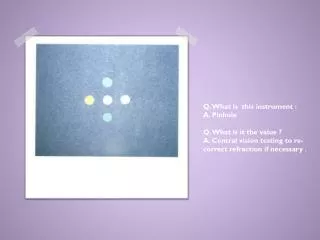
Q. What is this instrument : A. Pinhole Q. What is it the value ? A. Central vision testing to re-correct refraction if necessary .

E N D
Q. What is this instrument :A. PinholeQ. What is it the value ? A. Central vision testing to re-correct refraction if necessary .
Direct OpthlamoscopeQ. What is the magnification?A. x15 Q. Mention 2 characteristics of the image produced.A. Right image (not inverted), mono-ocular vision, high magnification, narrow area.
Q. Identify the organ (A)A. Superior canaliculus.Q. Identify the organ (B)A. Nasolacrimal sac. A B
1- Dx = nasolacrimal duct obstruction 2- Rx = recanalization or massage صورة طفل وضع له صبغة الاختبار وكأنه يبكي الصبغة ؟؟
1- INSTRUMENT ?Prism 2- value ?Test eye deviation (measure strabismus) إذا حابين تشوفونها تلقونها بالعيادات
Dx ?Neurofibromatosisassociated symptoms in eyes?-Sphenoidaldysplsia-Optic Glioma-Nearofibrosarcoma-Iris Hamartoma
Q. What is the diagnosis?Vitreous HemorrhageQ. Name 3 causes;Trauma, HTN , DM
Dx?Sublaxatedlens (SuperoTemporally)associated with ?→ Marfan’s Syndrome
Dx?Posterior synechiarisk factor?-trauma-iritis-iridocyclitis-syphilis-leprosy-herpes-toxoplasmosis-Parasites-tuberculosis-hypermature cataractTTT:AtropineMydriaticsTopical steroidsCAUTION ! NEVER give steroids if: (1) There are signs of infection. (2) He has a corneal ulcer.
Dx?Iridodialysiscauses:- Blunt & penetrating trauma- iatrogenic ( cataract surgery)TTT:-Patch & Observe-Surgical
This was a bilateral finding in a young obese woman with 120/80 BP. CT scan imaging was negative.Q. What is the most likely diagnosis?-papilledema-Brain tumor-Pseudotumorcrebri-HTNQ. How would you manage her?1.Medical: weight reduction & carbonic-anhydrase inhibitors (e.g. acetazolamide)2.Surgical: CSF shunt.
Dx?Pusdoesotropia.What is the cause?Large epicanthal fold.You can see the symmetrical light reflex
Dx?Rt eye exotropia.Type of surgery.Lateral rectus weakening.Medial rectus strengthening
Name the instrument?Exophthalmometer.uses ?Measure Exopthalmos
Q. What is the diagnosis?A. Right facial (7th) nerve palsy (LMNL).Q. Mention 2 ocular manifestations:Exposure keratitisepiphoria(excessive tearing)ectropion.
Q. What is the diagnosis?A. Accommodative esotropia in the right eye.Q. Which type of refractive error is associated with this condition?A. Hyperopia.
2 1 1- Ciliary Body 2- Central retinal artery
Q. DIAGNOSIS ?PROLIFERATIVE DIABETIC RETINOPATHYQ. What is the sign ?Fan shaped neovascularization on optic disc (NVD)Q. How would you manage this patient?A. Pan-retinal photocoagulation (PRP) & control blood sugar
Q. What is this sign?A. Leucokoria in the right eye.Q. Mention 2 differential diagnoses.-Congenital cataract-Retinoblastoma-Premature Retinopathy
Q. What is the diagnosis?A. Subconjuctival hemorrhage.Q. Mention 2 causes:-Trauma-blood diseases-anti-coagulants-OCP-cough-Valsalva maneuver-old age-idiopathic.
A patient with a history of glaucomaQ. Dx?A. Open angle glaucoma Q. What is this sign?A. Cupping (increased cup:disk).Q. Which type of visual field defect is associated with this condition?Peripheral visual field defectNasal StepArcuateScatoma
A 25 year old patient with a history of sinusitis &feverQ. What is the diagnosis?A. Orbital cellulitis.Q. How would you manage her?Admission, temperature chart, culture and sensitivity, IV antibiotics, CT scan.Q. SYMPTOMS ?- PROPTOSIS- RESTRICTED EYE MOVEMENT-Distorted vision-Optic nerve injuryComplications:Brain AbscessCavernous sinus thrombosisMeningitisOptic NeuritisVision loss
SLE butterfly rashRetinopath & KeratopathyCotton wool appearance
A patient with a history of wearing contact lensesQ. What is the diagnosis?A. Corneal ulcer.Q. How would you manage this patient?A. Remove the contact lenses & topical antibiotics
Q. What is the diagnosis?A. Herpitickeratitis.Q. What is the name of the stain that was used?A. Fluorescein dye.Q. Rx? acyclovir
ProptosisLid RetractionCauses:-Sinusitis-Trauma-Glucoma-Gravescomplications:Optic neuropathyRestricted eye movements
Q. What is the diagnosis?A. Senile cataract.Mention 2 postoperative complications:-Endophthalmitis-hemorrhage-IOL dislocation-Retinal Detachment-Cornea swelling-↑ IOP-Ptosis
A patient with a history of cataract surgeryQ. What is the diagnosis?Endophthalmitis.Q. How would you manage this patient? intravitrealantibiotics.
Q. What is this procedure called?A. Peripheral iridotomy.Q. What is the indication of this procedure?.-Acute closed angle glaucoma-narrow angle glaucoma.
Q. What is the diagnosis? A. Right oculomotor (3rd) nerve palsy. Q. If patient has a history of nausea, vomiting & dizziness. What will be the most likely diagnosis? Neoplasm (brain tumor).
Hx of corneal abrasion 1- Rx ? Patch & cover2- complication?-recurrent corneal erosions-Scar-Blepharospasm-Eye red-Eye pain-Photophobia مو نفس الصورة
HyphemaTTT: Bed rest & prevent bleedingCycloplegicsSteroidsAntifibrinolytics
Hx: Redness itching FB sensationAcute BlepharitisTTT: HygineAntibiotic ointmenthot compressor
Viteroushemorhage with Crescent shaped poolSymptoms: blurry vision• light flashes• floaters Causes:-DM-HTN-Sickle cell anemia-Carotid artery disease TTT: vitrectomy
CRAO-Cherry Red spot sign-Marcus Gunn PupilCauses:-HTN-DMGiant Cell Arteritis-Emboli-Carotid Artery disease-AtherosclerosisBlood diseasesTTT:-Restore blood flow in the 1st 2 days-supine position-ocular massage-IV acetazolamide& topical beta blockers
Q. What is the diagnosis?A. Central retinal vein obstruction (CRVO), sudden visual lossInvestigations: fluroscien angiographyQ. Mention 2 predisposing factors.-HTN & DM-Cardiovascular disorders-Bleeding or clotting disorders-Vasculitis-Autoimmune disorders-OCP-Trauma-Alcohol-Glucoma
1- Dx ?Foreign body in eye 2- Rx?- anesthesis- remove FB- topical antibiotics
1- Dx ?Herpes zoster ophthalmus(involving 5th nerve) Hutchinson’s sign (nasocilliary nerve)2- Rx?Acyclovir3- associated symptoms?-Headache-Fever-Malaise- eye pain-red eye (usually unilateral)-decreased vision-skin/eyelid rash-tearing
The Error : Myopia Correction by : Concave Lens or –velense
What is the refractive error illustrated in the diagram?A. HyperopiaQ. What type of lenses could be used to correct it?Convex lenses+ve lens
Q. What is the diagnosis?A. Keratoconus.Q. What is this sign?A. Munson’s sign.
Hx: glucome and medication that decrease iris pigmentation Prostaglandin Analogue