Structure and Function of the Ear
300 likes | 601 Views
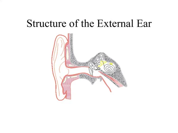
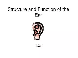
Structure and Function of the Ear. 1.3.1. 3 Sections to the Ear. External outer ear Middle Ear Inner Ear. External Ear. Pinna – visible part of the ear Also known as the auricle

Structure and Function of the Ear
E N D
Presentation Transcript
3 Sections to the Ear • External outer ear • Middle Ear • Inner Ear
External Ear • Pinna – visible part of the ear • Also known as the auricle • The function is to collect sound by acting as a funnel, amplifying the sound and directing it to the auditory canal The entire outer ear is the pinna. It is divided into different parts.
External Auditory Canal • From the external opening of the ear to the tympanic membrane (ear drum) • Lined with cells that secrete cerumen to protect the canal.
Tympanic Membrane – Ear Drum • A thin membrane that separates the external ear from the middle ear. • Its function is to transmit sound from the air to the ossicles inside the middle ear. • Vibrates when sound hits it
Middle Ear • Contain Ossicles – bony parts • 3 Main Bones - smallest bones in body • Malleus (hammer) • Incus (anvil) • Stapes (stirups) • Bones transfer sound coming off vibration of the tympanic membrane to the inner ear.
The Mastoid Process • Posterior portion of temporal bone • Projection behind ear • Attachment for certain neck muscles • Provides support for outer ear and inner ear
Eustachian Tubes • A tube that links the pharynx to the middle ear (mastoid). • The function of the Eustachian tube is to protect, aerate and drain the middle ear. • Maintains even air pressure around tympanic membrane
Inner Ear • Houses the sensory organs that help in hearing and maintaining balance.
Inner Ear • consists of a maze of fluid-filled tubes, running through the temporal bone of the skull. • The bony tubes, the bony labyrinth, are filled with a fluid called perilymph.
Inner Ear Parts • The front portion is the snail-shaped cochlea, which functions in hearing. • The rear part, the semicircular canals, helps maintain balance. • Interconnecting the cochlea and the semicircular canals is the vestibule, containing the sense organs responsible for balance, the utricle and saccule.
Cochlea • is the auditory portion of the inner ear. • Its core component is the Organ of Corti, the sensory organ of hearing, which is distributed along the partition separating fluid chambers in the coiled tapered tube of the cochlea.
The Organ of Corti • The end organ of hearing • Contains stereocilia and hair cells. • Respond to fluid-borne vibrations in cochlea • Structure that transduces pressure waves to action potantials • 15,000 to 20,000 auditory nerve cells with hair cells
Hair Cells • Frequency specific • High pitches= base of cochlea • Low pitches= apex of cochlea Sensory receptors of both the auditory system and the vestibular system in all vertebrates. Located in the Organ of Corti in cochlea of inner ear
Oval and Round Window • The oval window (or vestibular window) is a membrane-covered opening which leads from the middle ear to the vestibule of the inner ear. • As the oval window membrane moves in when hit by the stapes, the round window membrane moves out, and this allows movement of the fluid within the cochlea, leading to movement of the cochlear inner hair cells and thus hearing.
Cochlear Nerve • The cochlear nerve (also auditory or acoustic nerve) is a nerve in the head that carries signals from the cochlea of the inner ear to the brain. • The cochlear nerve is a sensory nerve, one which conducts to the brain information about the environment, in this case acoustic energy impinging on the tympanic membrane. • The cochlear nerve arises from within the cochlea and extends to the brainstem.
Vestibular Nerve • The vestibular nerve is one of the two branches of the vestibulocochlear nerve, functioning in tandem with the cochlear nerve. • Vestibular nerves have the job of transmitting data to and from the brain that have to do with the regulation of the sense of balance.
How Sound Travels Through The Ear... 1. Acoustic energy, in the form of sound waves, is channeled into the ear canal by the pinna 2. Sound waves hit the tympanic membrane and cause it to vibrate, like a drum, changing it into mechanical energy 3. The malleus, which is attached to the tympanic membrane, starts the ossicles into motion 4. The stapes moves in and out of the oval window of the cochlea creating a fluid motion 5. The fluid movement causes membranes in the Organ of Corti to shear against the hair cells 6. This creates an electrical signal which is sent up the Auditory Nerve to the brain The brain interprets it as sound!
What is letter A? • What is letter B? • What is letter D? • What is letter E and what is its function?
Answers • Outer ear • Middle ear • Pinna • Auditory canal – directs sound waves into the ear.