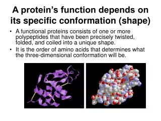
Function depends on structure
Function depends on structure. Muscle classification Striated muscle Skeletal Muscle - voluntary muscles that allow for movement Cardiac Muscle - heart - specialized, involuntary Non-striated muscle Smooth Muscle such as blood vessels, digestive tract, internal organs, involuntary.
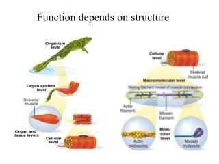

Function depends on structure
E N D
Presentation Transcript
Muscle classification • Striated muscle • Skeletal Muscle - voluntary muscles that allow for movement • Cardiac Muscle - heart - specialized, involuntary • Non-striated muscle • Smooth Muscle such as blood vessels, digestive tract, internal organs, involuntary
Muscle functions • Muscle perform four import functions: • Produce movement • Maintaining posture • Stabilizing joints • Generating heat
Functional characteristics of muscles Excitability (irritability): the ability to receive and respond to a stimulus Contractility: the ability to shorten forcibly when adequately stimulated Extensibility: the ability to be stretched or extended Elasticity: the ability of a muscle fiber to resume its resting length after being stretched.
Sarcomere: the contractile unit of a myofibril contains actin thin filament and myosin thick filament
I band A band I band Cross bridge Thick filament Thin filament Fig. 8-4, p.317
Contraction of sarcomeres-sliding-filament theory muscle contraction- sarcomeres shorten, actin and myosin move past each other and increase overlap between actin and myosin. muscle stretched- Sarcomeres elongate. Reduce overlap between actin and myosin. Note: Length of thick (myosin) and thin (actin) filaments remains constant.
Length-tension relation Total tension is proportional to the total number of cross-bridges (overlap) between actin and myosin filaments Ideal resting length: generate maximum force. Overlap to small: few cross-bridges can attach. No overlap: no cross-bridges can attach to actin.
3 pairs of molecules: • myosin heavy chains • essential light chains • regulatory light chains
Actin - thin filaments • 1. comprised of protein dimers linked in "chains"2. each actin monomer has a myosin binding site3. thin filaments are anchored at one end to "Z-line" proteins4. thin filaments are free at other end5. "sarcomere" is the name for unit between "Z-lines" G-actin polymerize F-actin (filamentous)
Troponin is a complex of 3 protein subunits: Troponin C binds Ca 2+ Troponin T binds tropomyosin Troponin I binds both actin & tropomyosin
Troponin C binds Ca 2+ Troponin T binds tropomyosin Troponin I binds both actin & tropomyosin
Cross-bridge chemistry • Attachable • Revisable Transduction of chemical to mechanical energy in muscle causes the filaments to slide: Partial rotation of the actin-bound myosin head.
neuromuscular junctions Each muscle cell is directly innervated by the terminal branch of a motor neuron. The contact between nerve and muscle occurs at a small specialized spot termed the neuromuscular junction (NMJ).
Transverse tube (T tube not Z disk): transmit excitation into muscle fibers Crab Frog
Sarcoplasmic reticulum (SR): Ca2+ is stored and released as free Ca2+ during excitation-contraction
Calsequestirin: Ca2+ binding protein in SR Ca2+/Ma2+ pump (ATPase): proteins in SR actively transport Ca2+ ions (requires ATP).
Ca++ regulation • a. neural activation >> muscle is electrically excited >> AP • AP ionic currents reach SR, open voltage sensitive Ca++ channelsCa++ rushes out of SR, binds to troponin C, actin-myosin permitted to interact >> contraction • b. AP stops, voltage sensitive Ca++ channels close, • Ca++ rapidly pumped into SR, tropomyosin returns, actin-myosin interactions blocked >> relaxation • c. Ca++ is sequestered (pumped and stored) in Sarcoplasmic Reticulum (SR) • - SR is the endoplasmic reticulum of muscle cells- SR is intracellular Ca++ store • d. Ca++ is actively pumped into SR from muscle cytoplasm
Ryanodine receptor: located on SR membrane • Dihydropyridine receptor: located on T tubule membrane, no or little Ca2+ passes through in skeletal muscle. • Releasing Ca2+ from SR into the myoplasm depends • interaction of activated dihydropyridine receptor and ryanodine receptor-plunger model • Calcium-induced calcium release
Mechanisms of Contraction • AP travels down the motor neuron to bouton. • VG Ca++ channels open, Ca++ diffuses into the bouton. • Ca++ binds to vesicles of NT. • ACh released into neuromuscular junction. • ACh binds onto receptor. • Chemical gated channel for Na+ and K+open.
Mechanisms of Contraction • Na+ diffuses into and K+ out of the membrane. • End-plate potential occurs (depolarization). • + ions are attracted to negative membrane. • If depolarization sufficient, threshold occurs, producing AP.
Mechanisms of Contraction • AP travels down sarcolema and T tubules. • Terminal cisternae release Ca++.
Mechanisms of Contraction • Ca++binds to troponin. • Troponin-tropomyosin complex moves. • Active binding site on actin disclosed.
Sliding Filament Theory • Sliding of filaments is produced by the actions of cross bridges. • Cross bridges are part of the myosin proteins that form arms that terminate in heads. • Each myosin head contains an ATP-binding site. • The myosin head functions as a myosin ATPase.
Contraction • Myosin binding site splits ATP to ADP and Pi. • ADP and Pi remain bound to myosin until myosin heads attach to actin. • Pi is released, causing the power stroke to occur.
Contraction • Power stroke pulls actin toward the center of the A band. • ADP is released, when myosin binds to a fresh ATP at the end of the power stroke. • Release of ADP upon binding to another ATP, causes the cross bridge bond to break. • Cross bridges detach, ready to bind again.
Contraction • ACh-esterase degrades ACh. • Ca++ pumped back into SR. • Choline recycled to make more ACh. • Only about 50% if cross bridges are attached at any given time. • Asynchronous action.
Contraction • A bands: • Move closer together. • Do not shorten. • I band: • Distance between A bands of successive sarcomeres. • Decrease in length. • Occurs because of sliding of thin filaments over and between thick filaments. • H band shortens. • Contains only thick filaments.
Regulation of Contraction • Regulation of cross-bridge attachment to actin due to: • Tropomyosin. • Troponin.
Role of Ca++ • Relaxation: • [Ca++ ] in sarcoplasm low when tropomyosin block attachment. • Ca++ is pumped back into the SR in the terminal cisternae. • Muscle relaxes.
Role of Ca++ in Muscle Contraction • Stimulated: • Ca++ is released from SR. • Ca++ attaches to troponin • Tropomyosin-troponin configuration change
Two major processes require ATP in muscle contraction: • Hydrolysis ATP by myosin (70-80%) • Pumping of Ca2+ back into SR (20-30%)