Download
1 / 45
450 likes | 474 Views
Learn about the importance of the skeletal, muscular, respiratory, and cardiovascular systems in physical activity. Discover how to design training programs, develop correct technique, minimize injury risk, and optimize performance.

E N D
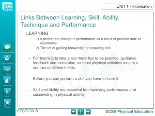
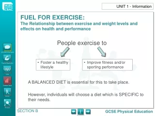
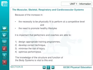
Because of the increase in: the necessity to be physically fit to perform at a competitive level and the need to promote healthy lifestyles it is important that performers and coaches are able to: design appropriate training programmes, develop correct technique, minimise the risk of injury, optimise performance. UNIT 1 - Information The knowledge of the structure and function of the Body Systems is vital to this end.
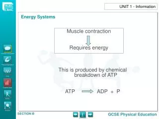
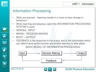
UNIT 1 - Information The skeletal and muscular systems work together to allow movement for physical activity. A The respiratory and cardiovascular systems work together to supply oxygen/ energy for muscle contraction. B
There are 206 bones in a human skeleton cranium clavicle mandible scapula sternum rib humerus vertebra radius pelvis ulna sacrum cocyx carpals metacarpals phalanges femur patella tibia fibula tarsals metatarsals UNIT 1 - Information The Muscular, Skeletal, Respiratory and Cardiovascular Systems Show / hide bone names Show / hide arrows
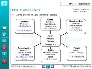
The skeleton has many FUNCTIONS when taking part in physical activity. These include: Providing SUPPORT for the movement taking place. PROTECTING vital organs against impact and injury. Producing RED BLOOD CELLS which help carry O2 (Oxygen) for exercise. Providing points for MUSCULAR ATTACHMENT – ORIGIN/ INSERTION. Allowing MOVEMENT through the JOINT and LEVER systems. UNIT 1 - Information GCSE Physical Education
UNIT 1 - Information IRREGULAR (Provides support and protection) e.g. vertebrae LONG (Provides effective levers for movement) e.g. femur SHORT (Well adapted to resist compression forces and help in performing fine movements) e.g. carpals BONES FLAT (Protective function) e.g. sternum
UNIT 1 - Information APPENDICULAR Arms, shoulders, hips, legs SKELETON Skull, spine, sternum, ribs AXIAL
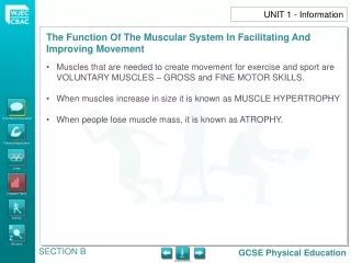
Physical activity stimulates the production of bone tissue to compensate for the stresses of training. If the intensity is too high, damage and injury of the bone can occur e.g. shin splints. Reduced stress results in bone weight and strength reduction – ATROPHY – usually after a lay-off due to injury. It is important to return to full training slowly, to allow the bones to adapt. UNIT 1 - Information
JOINTS Joints enable the body to move. Joints are the place where 2 bones meet. Each type of physical activity will make particular demands on certain joints e.g. butterfly swimmers require a wider than natural range of movement at the shoulder joint – ball and socket. Joints are structured for either STRENGTH or MOBILITY: UNIT 1 - Information SHOULDER JOINT Shallow cavity to allow more movement but is less stable HIP JOINT Supports body weight – is more stable – less movement allowed
Good joint mobility is often an essential requirement to good performance in physical activity and has implications regarding training programmes. Joints can be classified according to the amount of movement they allow: UNIT 1 - Information 1) IMMOVABLE / FIBROUS JOINTS These joints are fixed and no movement is possible. Examples are found in the skull and pelvic girdle – areas of the body where great strength is required. 2) SLIGHTLY MOVABLE / CARTILAGINOUS JOINTS There are small gaps between the bones at these joints, filled with CARTILAGE to prevent grinding and enable slight movement. Examples are found between the vertebrae and between the ribs and sternum. 3) FREELY MOVABLE / SYNOVIAL JOINTS
The majority of joints in the body are synovial – they allow the greatest range of movement. The bones are linked by LIGAMENTS. There are 6 types of synovial joint, and each type has its own characteristic range and type of movement. They are designed to stop and prevent friction between the moving bones. Examples are the shoulder, elbow, hip, knee and ankle joints. These joints are important for physical activity – they are often put under a great deal of stress (e.g. the knee), but are so designed to be robust and withstand pressure. UNIT 1 - Information
TYPE OF MOVEMENT FLEXION – Reducing the angle at a joint (bending). UNIT 1 - Information
TYPE OF MOVEMENT EXTENSION – Increasing the angle at a joint (straightening). UNIT 1 - Information
TYPE OF MOVEMENT CIRCUMDUCTION – A circular movement, which combines flexion, extension, abduction, and adduction so that the movement of the body-part describes a cone shape. UNIT 1 - Information
TYPE OF MOVEMENT ROTATION – Is a circular movement made by a joint. UNIT 1 - Information
TYPE OF MOVEMENT ABDUCTION – Is the sideways movement at the hip and shoulder joints away from the body. UNIT 1 - Information
TYPE OF MOVEMENT ADDUCTION – Is movement at the hip and shoulder joints towards the body. UNIT 1 - Information
UNIT 1 - Information SECTION B
UNIT 1 – Information Ball and socket
UNIT 1 - Information Hinge Hinge
UNIT 1 - Information Pivot Atlas Vertebrae Axis
UNIT 1 - Information Condyloid Condyloid joint Movement GCSE Physical Education
UNIT 1 - Information The Muscular, Skeletal, Respiratory and Cardiovascular Systems Saddle Saddle joint
UNIT 1 - Information Gliding Gliding occurs between the small bones
Strong joints are essential for: Coping with stress of physical activity. Contributing to a healthy, active lifestyle. Making skilled movements. UNIT 1 - Information FUNCTIONS OF LIGAMENTS, TENDONS AND CARTILAGE Ligaments, tendons and cartilage provide the strength, mobility and protection needed to help the joints, increase the flexibility and provide a greater range of movement.
Ligaments are strong, fibrous, non-elastic bands of tissue which attach bone to bone across a joint. They control the movement and the stability of the joint. They hold a joint in place. They prevent unwanted movements at a joint. UNIT 1 - Information LIGAMENTS Information/Discussion CARTILAGE Practical Application • Cartilage protects bone. • It is a tough, smooth tissue that covers the end of bones and acts as a shock absorber. • It reduces friction between the bones. • Cartilage damage may occur over a period of time because of the constant stress/ impact and twisting/ turning of physical activity. Links Diagram/Table Activity Glossary GCSE Physical Education SECTION B
Tendons attach MUSCLE to BONE. They are strong but flexible. When muscles contract (shorten), they exert a pull on that tendon which acts across the joint to make a joint move. UNIT 1 - Information TENDONS GCSE Physical Education
UNIT 1 - Information The Muscular, Skeletal, Respiratory and Cardiovascular Systems The structure of a knee joint showing the bones, ligaments, tendons, muscle and cartilage involved Tendon (this joins muscle to bone) Muscle Ball-shaped end to long bone of leg or femur (rounded ends to bones ensure easy movement with little friction) Patella (kneecap) Synovial fluid (oils or lubricates the joint helping it to move more easily) Cartilage (smooth, slippery, reduces friction, helps to reduce shock) Show Bone Show Muscle Ligament (tough strip of tissue joining bone to bone) Show Tendons Tibia Show Ligament Show Cartilage GCSE Physical Education
1. Match the scientific name for the bones on the left of the table with the common names by dragging them to the table. You can also drag the numbers to the diagram of the skeleton. UNIT 1 – Practical Application 1 1 2 2 3 3 4 4 5 5 THIGH BONE FINGERS/TOES HIP SKULL JAW BONE
2. Class activity: e.g. Set shot – Basketball (Teach and class practice) a) Attempt to break the skill down into ‘PHASES’. b) Construct a table, and for each phase: (i) identify the moving joint; (ii) name the type of movement; (iii) identify the muscles that are at work. (For (iii), remind groups that muscles can only PULL. Also, a muscle must span a joint if it is to move it – ORIGINS/ INSERTIONS). UNIT 1 – Practical Application
3. Class activity: e.g. Hockey – group to identify the function of the following structures in a warm-up: Synovial fluid; Ligaments; Articular cartilage. UNIT 1 – Practical Application 4. Class to identify which muscles are agonists/ antagonists for the following joint actions during the ‘PUSH’ in hockey: Extending the knee. Flexing the elbow.
5. In a game of hockey e.g. identify when a player would use: a) mainly FAST TWITCH fibres; b) mainly SLOW TWITCH fibres. Do different positions in a game of hockey place different demands on muscle fibre demand, and if so, why? UNIT 1 – Practical Application Ball Games: Joint Movement 6. Work with a partner and identify basic body movements, joints and analysis of specific movements from chosen activities. 7. Consider the factors affecting performance and participation e.g. age and range of movement around a joint. 8. Identify the joints involved in, for example, throwing a ball (netball). 9. Identify the joints involved in, for example, kicking a ball (football). 10. Discuss the different types of movement around a joint. 11. Develop by pupils choosing their own activity and identifying movements at joints. 12. The above links with ‘specific’ training sessions and ‘specific’ training methods for sporting activity.
UNIT 1 – Practical Application Analysis of Movement: 13. In pairs, discuss basic movements and actions of joints. 14. Activity and sport specific discussion regarding movement, muscles and joint actions. 15. Record movements and use for analysis/ discussion. 16. Consider the link between desired movement patterns, skill and sport specific fitness training.
Muscular system Respiratory system Cardiovascular system Aerobic/ Anaerobic systems Energy continuum Training zones Intensity/ duration of exercise Short-term effects of exercise on the systems of the body Long-term benefits of exercise on the systems of the body Body types UNIT 1 - Links The Muscular, Skeletal, Respiratory and Cardiovascular Systems
UNIT 1 - Activity The Muscular, Skeletal, Respiratory and Cardiovascular Systems 1. “Muscles and joints work together to produce and control movement”. • In the diagram below, which muscle contracts to cause the bending of the knee? Is it: a) the quadriceps or b) the hamstring? • What type of synovial joint is the knee joint and what type of movement does it allow? (iii) What attaches bone to a bone to ensure stability of the joint? A B
UNIT 1 - Activity 2. The diagram below shows the action of the upper arm muscle involved in lifting a weight. Study the diagrams and then answer the questions below. Diagram 1 Diagram 2 Biceps A Biceps A Triceps B Triceps B • In diagram 1, what is the movement upwards called? • Which muscle is shortening (contracting) to cause this upward movement, is it A or B? • If the contracting muscle is the agonistic or prime mover, what is the relaxing (flexing) muscle called? • In diagram 2, what is the downward movement called? • What attaches the muscle to a bone to help in movement?
UNIT 1 - Activity 3. “Good joint mobility is essential for efficient performance in most sporting activities.” Complete the following table: (iii) Describe an example of a movement in sport which involves (a) flexion/ extension and (b) adduction/ abduction.
B Hamstring group Quadriceps group A Gastrocnemius Ball UNIT 1 - Activity 4. The diagram below shows the leg of a football player preparing to kick a ball. • Which PRIME MOVER (AGONIST) muscle would need to CONTRACT (FLEX) in order to LIFT the lower leg in readiness to kick the ball? • Which PRIME MOVER (AGONIST) muscle would need to CONTRACT (FLEX) in order to KICK the ball? • Identify the type of joint at the KNEE (A). • Which joint, A or B allows most movement?
B Hamstring group Quadriceps group A Gastrocnemius Ball UNIT 1 - Activity • What is the term used for muscles which RELAX to allow movement to take place? • What attaches muscle to bone in order for movement at the joint to take place? • What attaches bone to bone to give joints stability? • The movement in the diagram is an example of which type of muscular contraction?
A B Diagram 1 Bend Diagram 2 Jump UNIT 1 - Activity 5. The diagrams below shows the action of the leg muscle and joints involved in a STANDING LONG JUMP. Study the diagrams and then answer the questions below by completing the second column in the table. Show action
UNIT 1 - Activity Muscles act in pairs, some contract while others extend. Revisit diagram
UNIT 1 - Activity 6. Describe the movement at each joint Using a grid like the one above, describe the type of movement at each position while: ● Running ● Diving – racing – front crawl ● Push pass (hockey) ● Any other activity of your choice
BONE BONE UNIT 1 - Activity 7. Label the diagram of a typical joint shown below by dragging the labels to their appropriate places Joint capsule – contains and protects the joint structures. Articular cartilage – protects ends of bones by acting as a shock absorber and reducing friction. Synovial membrane – secretes synovial fluid. Synovial fluid – lubricates the joint, helping the bones move more easily. Ligaments – strong strap-like structures which prevent too much sideways movement.
UNIT 1 - Activity • Explain, using examples from sport, what the different categories of joints are and why we need them. 9. Complete the following table about the main categories of joints. Sutures of skull Synovial (freely movable) Some movement Slightly moveable Hip, shoulder, knee, elbow Very little movement 10. What factors affect the amount of movement possible at a joint?
UNIT 1 - Activity 11. The diagram below shows the action of the upper muscle involved in lifting a weight. Study the diagrams and then answer the questions below. • Which muscle is involved in bending the arm? • Which muscle is involved in straightening the arm? • Which type of movement is taking place at the elbow joint? • This movement is an example of a third order lever in action. Identify the pivot/ fulcrum and load/ resistance by dragging the labels to the appropriate points. Biceps A Triceps B Pivot Load
UNIT 1 - Activity 12. • Explain why synovial joints are so important for the sportsperson. • Explain why it is important to train all the muscles acting on a joint. • “Different synovial joints allow certain types of movement”. Complete the table for both extension/ flexion and rotation.