Download
1 / 96
970 likes | 1.06k Views
Learn about granulomatous diseases affecting the nose and paranasal sinuses, including tuberculosis and leprosy. Explore diagnostic features, symptoms, treatment options, and complications associated with these conditions.

E N D
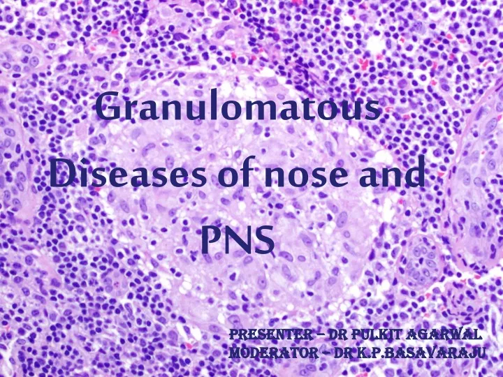
Granulomatous Diseases of nose and PNS Presenter – Dr Pulkit Agarwal Moderator – Dr K.P.Basavaraju
What is Granuloma ? • Immune system tries to wall off foreign substances but fails • Bacteria, Fungi, keratin, suture material. • Histopathologically – Chronic inflammation • Epitheloid cells • Multinucleate giant cells • Lymphocytes & fibroblasts • Necrosis and Vasculitis.
Granuloma as misnomer • “Granuloma" loosely to mean "a small nodule". • Vocal cord granuloma • Pyogenic granuloma • Intubation granuloma • Pulmonary hyalinizing granuloma i.e. keloid-like fibrosis in the lung • Radiologists - calcified nodule on X-ray or CT scan of the chest. • Most accurate use – Pathologists who observe stained tissue under microscope. Granulation tissue
Tuberculosis : • Nasal Mucosa involvement rare. • Portal – Inhalation (commonest), ingestion, inoculation. • Asso with Primary pulm. Tuberculosis • M. tuberculosis and M. bovis - main pathogens. • Most common affection of nose – Lupus Vulgaris • Macroscopic appearance: • Nodular form (Lupus) • Ulcerative form • Sinus granuloma
Indolent and chronic from of tb infection affecting the skin and mucous membrane of nose. • F:M = 2:1 (young adults) • Temperate Climate disease • Starts at Vestibule, extends to adjoining skin and mucosa with mucocutaneous junction as MC location. • Pathogenesis – Direct dermal inoculation (patient having immunity to infection due to previous exposure)
Scarring of Vestibule common in later stages • HPE – Shows tuberculous granulomas, RE cells at centre which soon necrose & coalesce, surrounded by ring of living RE cells. • Around it is ring of Lymphocytes, plasma cells & fibroblasts. • Multinucleate giant cells scattered throughout
Symptoms : • Nasal discharge • Nasal obstruction • Presence of non foul smelling crusts • Epistaxis • Evening rise of temperature • Mild fetor if ulcerated • Pain is unusual • Apple jelly nodules (Early lesions) - reddish firm nodules at mucocutaneous junction of nasal septum. • May later coaslesce & break down to form characteristic ulcers with a pale granular base and undermined edges
Diagnostic Features : • Blanching (glass slide) • Bacterial examination • Biopsy (diagnostic) • Culture - Lowenstein-Jensen slope • Recent - BACTEC
Complications: • Pulmonary tuberculosis • Corneal ulceration • Dacryocystitis • Nasopharyngeal lupus • Lupus of face • Atrophic rhinitis • Development of epithelioma (rare sequlae) • Sudden increase in size, hardness of the node in an elderly with increasing tendency to bleed should arouse the suspicion of epithelioma formation.
Ulcerative Form • Ulcers are commonly found on the anterior part of the cartilagenous nasal septum, inferior turbinate and anterior choanae. • Ulceration Fibrosis Distortion of Nasal Alae • If turbinates are involved extensively, the lining ciliated columnar epithelium is not renewed leading to secondary atrophic rhinitis. • Spread usually occurring in a backward direction from the primary site • Only cartilagenous septum involvement so no sinking of nasal bridge.
Sinus Granuloma • Isolated sinus involvement may occur without any signs or symptoms in the nose. • Usually presented with a diffuse soft tissue swelling and multiple discharging sinuses in the supraorbital region secondary to osteomyelitic involvement of the frontal bone. • The computed tomography (CT) and magnetic resonance imaging (MRI) characteristics are nonspecific and demonstrate a soft tissue mass with or without bone destruction. • Inv. of the orbit & its nerves may occur. • Diagnosis by an external or endoscopic biopsy.
Specific Investigations • Montaux test • AMTD (Amplified Mycobacterium Tuberculosis Detection) • PCR • Fluorescence microscopy
Treatment • Antituberculous drugs • Short Course Chemotherapy : • Extends over 6-9 months and consists of Rifampicin, INH, Ethambutol and Pyrizinamide • DOTS : • Initial Phase - 2 mnths - HRZE • Rifampicin (R) - 450mg • INH (H) - 600mg • Ethambutol (E) - 1200mg • Pyrizinamide (Z) - 1500mg
Treatment • Maintainence treatment (4 months) - (HR)3 (thrice weekly under observation) • Vit D supplementation - hastens sputum culture conversion (only in vit D deficient cases) • Surgical correction
Leprosy • Caused by M. Leprae which is morphologically similar to M. Tuberculi • Non culturable, inoculated into experimental animals • Based on the host's immunological status and clinical, histological and microbiological features leprosy divided into types.
Types of leprosy : Tuberculoid leprosy: • Immunocompetent patients • Solitary lesions (anesthetic patches) • Involvement of one / more related sensory or motor nerves. • Nasal Vestibule involved without mucosa involvement • Isolated cranial nerve palsies (V and VIIth) have also been documented
Lepromatous Leprosy : • Least immunological resistance to the organism. • Diffuse infiltration of skin, nerves and mucosal surfaces. • Cutaneous infiltration is more common over the edges of pinna, chin, nose and eyebrows. • Highly infectious nasal discharge.
Signs and Symptoms • Earliest sign - nodular thickening of nasal mucosa • Crust formation • Nasal obstruction (out of proportion) • Sero sanguineous discharge Nodules are paler than the surrounding mucosa with a yellowish tinge. Commonly begin at the anterior end of inferior turbinate, progresses to gross inflammation of nasal mucosa.
Both the bony and cartilaginous portions of nasal septum are destroyed due to perichondritis and periosteitis (nasal bridge collapse) • Anterior nasal spine destruction seen • Radiographs showing the absence / erosion of anterior nasal spine are virtually diagnostic of Lepromatous leprosy • Hyposmia is seen in 40% • Nasal mucosa scraping microscopy shows typical cigar pattern Lepra bacilli
Borderline leprosy • Poor immunological resistance • May progress to lepromatous leprosy • Skin lesions are more numerous and are seen around eyes, nose and mouth • In pure borderline lesions - nasal mucosa involvement is not seen • Nasal mucosal inv suggests progression to LL.
Diagnosis • Early diagnosis is essential since nasal discharge is principle route of transmission of the disease • Clinical findings of anaes. on the skin, thickened peripheral nerves and skin lesions, & supplemented by bacteriological examination. • Confirmed by dem. of M. leprae on microscopy of the nasal discharge or scrapings of nasal mucosa, or histopathology of nasal mucosa. • Skin biopsy in tuberculoid leprosy
Treatment • Dapsone reduces the bacilli count of nasal discharge to zero or near zero within a couple of months BUT increased resistance • Modern drugs like Rifampicin and Clofazimine can reduce the bacilli count to zero within 10 days • Triple therapy: • Rifampicin – 600 mg on first two days of a month taken before breakfast • Clofazimine – 100 mg on alternate days for three times a week • Dapsone – 100 mg a day
Rifampicin stopped after 3 mnths • Rest 2 continued till 9 mnths • Intranasal Rifampicin much faster than oral route • Nasal douching for removal of crusts • Betnovate (1 part) in Unguentum (2 parts) has been used with good results
syphilis • Nasal syphilis an occur at any age • Uncommon as signs symptoms difficult to elicit in early stages especially if antibiotics given • Causative org. – Treponema Pallidum(Spirochete) • Pathogenesis - The organisms usually reside and multiply in the perivascular lymphatics of the blood vessel wall. • Local host reaction results in an infiltration by polymorphs, activated lymphocytes (CD4+ and CD8+), plasma cells and histiocytes which are typical of syphilis.
The resultant endarteritis of small blood vessels with secondary hypertrophic changes in the endothelium, may lead to endarteritis obliterans and luminal obliteration. • HPE - Diagnosis is purely histopathological • Characterized by oedema, stromal infiltration with lymphocytes, plasma cells and endothelial cells • Perivascular cuffing by these cells and endarteritis will cause a reduction in the lumen of blood vessels causing necrosis and ulceration
Nasal syphilis can be classified into: • Primary syphilis • Secondary syphilis • Tertiary syphilis • Primary syphilis : • Also known as Chancre, seen at external nose or inside the vestibule. • Appears as a hard, non painful ulcerated papule always associated with enlarged, rubbery, and non tender lymphadenopathy
A sore or chancre develops at the site of inoculation 10-90 days (average 21 days) following infection • These lesions undergo spontaneous resolution within 6-10 weeks • Should be differentiated from malignant neoplasms and furunculosis • Malignant neoplasms – affect older ages, relentless progression • Furunculosis – Painful condition, progresses to suppuration.
Treatment • Anti syphilitic Rx - i.m. penicillin. • The chancre may be cleansed with 1:2000 solution of per chloride of mercury and the surface smeared with 2% yellow mercuric oxide ointment. • The following points should be borne in mind : • Cultures from the surface of the lesion will always be negative
Smears when examined under dark ground illumination will show the spirochete Treponema palladium • Serological tests for syphilis may be positive – VDRL, TPHA, FTA-ABS. (The VDRL is a nontreponemal test and is relatively nonspecific but becomes positive early (four to five weeks) and correlates well with disease activity.) • A biopsy from suspicious lesion may confirm the diagnosis
Secondary Syphilis • Most infectious of all the three stages • Symptoms 6-10 weeks after inoculation • Secondary syphilis is rarely recognized in the nose, as mucous patches hardly ever occur on such a thin, attenuated mucous membrane.
The symptoms include: • Simple catarrhal rhinitis (persistent) • Crusting & fissuring of nasal vestibule • Other secondary lesions like mucous patches in the pharynx are also seen • Roseolar / papular skin rashes • Shotty enlarged non tender lymph nodes. • The scar of the primary lesion in the genitals or elsewhere may be visible.
Serological tests for syphilis are positive. • Dark-field examination of moist or intentionally abraided dry lesions may demonstrate the organism. • Lymph node aspirates or tissue specimens (lymph nodes, liver, skin or mucous membrane) may also be examined by silver stains or immunofluorescence for detection of spirochaetes • These patients respond to antisyphilitic drugs.
Tertiary Syphilis : • Only 1/3rd of secondary syphilis cases progress to show clinical manifestations of tertiary syphilis. • This stage is commonly encountered in the nose. • Lesion is also known as gumma. • This lesion invades the mucous membrane, periosteum and bone. • The bony portion of the nasal septum is frequently affected causing septal perforation.
Rarely the following portions of the nose can also be involved : • Lateral nasal wall • Frontal sinus • Nasal bones • Floor of the nose
Symptoms include: 1. Pain / headache (worse during night) 2. Swelling / obstruction of nose - swelling may be diffuse / localized associated with offensive discharge, bleeding and crusting of the nose 3. Olfactory acuity diminishes 4. Perforation of bony portion of nasal septum associated with collapse of the bridge of the nose can cause structural damage to the nasal architecture 5. There may also be associated secondary atrophic rhinitis
Nasal complications of gumma: 1. Secondary infections with pyogenic organisms 2. Sequestration 3. Perforation of bony portion of nasal septum, palate or nasal walls 4. Collapse of bridge of nose with deformity of nose 5. Scarring / stenosis of nasal passages 6. Atrophic rhinitis 7.Intracranial complications due to involvement of meninges
Treatment : • DOC for all stages – parentral Penicillin • Alkaline nasal douches one to three times a day. • Local yellow mercury oxide ointment. Dilute mercuric nitrate ointment should be applied freely to the nasal vestibules. • Atrophic rhinitis and deformity may persist after the disease is cured and this may need further surgery.
Congenital syphilis “Snuffles”: : • Any of the lesions of secondary and tertiary forms of syphilis of nose can occur. • Classically begin during the 3rd week of life. • May appear around 3rd month also • At First – simple catarrhal rhinitis. • Later becomes purulent with sec. fissuring and excoriation of the nasal vestibule and upper lip.
Gummatous and destructive lesions occur most commonly at puberty. • Mucous membrane, periosteum and bone may all be affected. • Other stigmata of syphilis, particularly Hutchinson's incisors and Moon's molars, interstitial keratitis, corneal opacities and sensorineural deafness may be present, and there may be a family history of syphilis, miscarriages and still births. • Serologic tests for syphilis will invariably be negative
Treatment : • In snuffles, the airway must be restored for suckling • The nasal discharge is removed by gentle suction and irrigation and the use of local drops of 0.5 percent ephedrine solution or simple normal saline, and by hyper extending the head before feeding • In the tertiary forms, simple nasal toilet by syringing with isotonic alkaline douche solution will remove the crusts and discharge, and yellow mercuric oxide ointment may be applied frequently to the nasal vestibules
Yaws (framboesia) • Closely resembles syphilis in its pathology. • Causative organism : Treponema Pertenue • Morphologically this organism is indistinguishable from Treponema pallidum. • Transmission: Is by direct extra genital contact. It has a high incidence in infancy and childhood. • Clinical features : • Primary, secondary and tertiary stages occur as in syphilis • Yaws principally affects the skin.
Mucous membrane are usually spared but for the mucocutaneous junctions. • Nasal lesions are very rare and do not differ in appearance from that of syphilis. • Advance nasal lesions are associated with extensive destruction of the nose, palate. • Destruction also may involve the whole of the maxilla, face and pharynx. • Serologic tests for syphilis is positive. • These lesions characteristically respond to conventional antisyphilitic treatment.
Rhinoscleroma (scleroma) • Progressive granulomatous disease commencing in the nose and later extending into the nasopharynx and oropharynx, the larynx and sometimes the trachea and bronchi. • Laryngeal involvement may occur in almost half the cases and the disease is perhaps better designated as respiratory scleroma, rather than rhinoscleroma. • Aetiology is multifactorial and usually seen in low socio economic status and poor domestic hygiene
Pathology : • Caused by gram negative bacilli Klebsiella rhinoscleromatis / Frisch bacilli / diplo bacillus. • F>M, any age • This organism could be a secondary invader following a viral infection. • Resides intracellularly and can be difficult to isolate in the laboratory. • The characteristic histological features include granulomatous tissue infiltrates in the submucosa, characterized by the presence of plasma cells, lymphocytes and eosinophils.
Besides, there are scattered large foam cells (Mikulicz cells) which have a central nucleus and a vacuolated cytoplasm containing frisch bacilli and Russel bodies. • The Mikulicz cells are transformed macro phages which have ingested the bacillus, but the bacillus being resistant to digestion by the macrophage persists intracellularly. • The Russel bodies resemble plasma cells with an eccentric nucleus and deep eosin staining cytoplasm. • Histochemical studies have indicated a high content of mucopolysaccharides around the walls of the bacillus • it is hypothesized that this may be responsible for the protection afforded to the organism from antibiotics and the hosts antibodies