Download
1 / 26
260 likes | 354 Views
Learn about the intricate pathways of taste and vision in human anatomy, including the role of facial, glossopharyngeal, and vagus nerves in taste perception, and the accessory structures of the eye that protect and maintain vision health. Understand the sensitivity of taste when linked with smell, and discover how the eye's structures like palpebrae, eyelashes, and lacrimal apparatus work together to ensure proper function and protection.

E N D
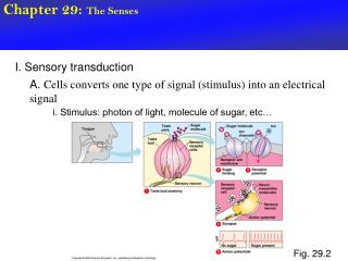
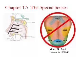
Taste Pathways • Facial, Glossopharyngeal and Vagus • Trigeminal—gives us “peppery and spicy hot” • Several thousand times more sensitive to taste when smell is involved
Vision • Accessory Structures • Palpebrae—Eye lids • Lubricate • Free from dust • Protect
Accessory cont… • Eyelashes • Protections
Accessory cont… • Sebaceous Glands • In eyelashes • Keeps eyelids from sticking • Sty—Painful swelling • Due to bacterial infection
Accessory cont… • Conjunctiva • Membrane over eye • Very sensitive • Conjunctivitis • AKA: Pinkeye • Damage/irritation to conjunctiva
Accessory cont… • Lacrimal Apparatus • Produces, distributes and removes tears • Constantly “crying” • Moistens and cleans • Reduce friction • Remove debris • Prevent bacterial infection • Provide nutrients and oxygen to conjunctiva
Accessory cont… • Nasolacrimal duct • Connects eye to nose
Accessory cont… • Extrinsic eye muscles • Control eye position
The Eye • Fibrous Tunics • Outermost layer of the eye • Support and protection • Attachment site for muscles • Assists in focusing
Fibrous tunics cont… • Sclera • White of the eye • Muscles insert here
Fibrous cont… • Cornea • No blood vessels • Limited repair
Vascular tunics • Blood vessels for the eye • Regulates light into eye • Controls shape of lens