Download
1 / 45
460 likes | 844 Views
Skeletal Muscle: Structure & Function Dr. Kyle Coffey. Week 7&8. Skeletal Muscle. Human body contains over 400 skeletal muscles 40-50% of total body weight Functions of skeletal muscle Force production for locomotion and breathing Force production for postural support

E N D
Skeletal Muscle: Structure & FunctionDr. Kyle Coffey Week 7&8
Skeletal Muscle • Human body contains over 400 skeletal muscles • 40-50% of total body weight • Functions of skeletal muscle • Force production for locomotion and breathing • Force production for postural support • Heat production during cold stress
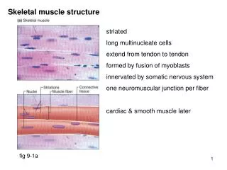
Structure: Connective Tissue • Epimysium • Outermost; surrounds entire muscle • Perimysium • Surrounds bundles of muscle fibers • Fascicles • Endomysium • Surrounds individual muscle fibers • Sarcolemma • Muscle cell membrane
Muscle Cells • Multi-nucleic • Allow for greater protein synthesis • Striated appearance, alternating light and dark bands • Dark bands contain contractile proteins • Satellite cells: undifferentiated cells that play role in muscle growth and repair • Increase number of nuclei during/after strength training
Microstructure of Muscle Fibers • Sarcomere: basic unit of muscle (“Z-line to Z-line”) • Z line (divide sarcomeres) • M line • H zone (myosin without actin overlap) • A band (myosin) • I band (actin) With contraction, what do you think occurs with the Z-lines?
Microstructure of Muscle Fibers • Myofibrils • Threadlike structures that contain contractile proteins • Actin (thin filament) • Myosin (thick filament) • Tropomyosin • Part of actin that contains attachment site for myosin • Troponin • Attached to tropomyosin • Covers attachment site for myosin
Microstructure of Muscle Fibers • Sarcoplasmic reticulum:pumps Ca+ ions • Surrounds each myofibril and runs parallel to it • Terminal cisternae: Storage sites for calcium • Transverse tubules • Invaginations in sarcolemma (membrane) • Linked closely to sarcoplasmic reticulum and terminal cristae • Allows quick depolarization of the inside of muscle cell
Neuromuscular Junction (NMJ) • Junction between motor neuron and muscle fiber • Motor unit • Motor neuron and all fibers it innervates • Motor end plate • Pocket formed around motor neuron by sarcolemma • Neuromuscular or Synaptic cleft • Short gap between neuron and muscle fiber • Acetylcholine is released from the motor neuron • Causes an end-plate potential (EPP) • Depolarization of muscle fiber
The Sliding Filament Theory • Contraction is considered excitation-coupling • Muscle shortening occurs due to the movement of the actin filament over the myosin filament • Formation of cross-bridges between actin and myosin filaments • Muscle shortening: reduction in distance from Z-line to Z-line • “Power stroke” • Generation of force
Sarcomere Shortening during Muscle Contraction How can we use this graphic to better understand active and passive insufficiency?
Power Stroke • Think of a crew team with synchronized oar strokes • Shorten muscle fiber by approximately 1% • Many muscles can shorten up to 60% of its resting length • Clear that contraction cycle can be repeated over and over
Energy for Contraction • ATP is required for muscle contraction • Myosin ATPase breaks down ATP as fiber contracts • Enzyme located on head of myosin cross bridge • ATP ADP + Pi • Sources of ATP • Phosphocreatine (PC) • Glycolysis • Oxidative phosphorylation So…is ATP needed in order for the actin/myosin cross bridge to form or separate?
Excitation-Contraction Coupling • Depolarization of motor end plate (excitation) is coupled to muscular contraction • Acetylcholine binds to motor end plate first • Action potential travels down transverse tubules and causes release of Ca+2 from SR • Ca+2 binds to troponin and causes positional change in tropomyosin • Exposing active sites on actin • Strong binding state formed between actin and myosin cross bridges • Contraction occurs
The Relationships Among Troponin, Tropomyosin, Myosin, and Calcium
Thoughts… • What is needed for a contraction cycle to be continued indefinitely? • What is the signal to stop a contraction? • How are troponin and tropomyosin regulators of muscle contraction?
Step-by-Step Summary of Excitation-Contraction Coupling • Excitation • Action potential in motor neuron causes release of acetylcholine from nerve fiber branches into synaptic cleftof neuromuscular junction. • Acetylcholine binds to receptors on motor end plate of sarcolemma • This binding leads to depolarization that is conducted down transverse tubules. • This causes release of Ca+2 from sarcoplasmic reticulum (SR). SR runs parallel to the myofibrils.
Step-by-Step Summary of Excitation-Contraction Coupling • Contraction • At rest, myosin cross-bridges in weak binding state (allows muscles to stretch quite easily). • Ca+2 binds to troponin, causes shift in tropomyosin to uncover active sites on actin • As myosin heads move to bind to actin, Pi releases; the resultant connection is called a cross bridge. • Cross-bridge movement or “power stroke” occursand ADP is released from myosin. • ATP attaches to myosin, breaking the cross-bridge and forming weak binding state, also called recovery stroke. • Then ATP binds to myosin, broken down to ADP+Pi, which energizes myosin.
Video • Sliding Filament Theory • Myosin and Actin
Muscle Fatigue • Decline in muscle power output due to: • Decrease in force generation • Decrease in shortening velocity • High-intensity exercise (~60 seconds) • Accumulation of lactate, H+, ADP, Pi, and free radicals • Diminishes cross bridges bound to actin and thus force production • Long-duration exercise (2–4 hours) • Muscle factors • Accumulation of free radicals • Electrolyte imbalance • Glycogen depletion What factors effect the fatigue process?
Exercise-Induced Muscle Cramps • Spasmodic, involuntary muscle contractions • Multiple theories • Electrolyte depletion and dehydration theory • Water and sodium loss via sweating causes spontaneous muscle contractions (motor nerve terminals spontaneously discharge) • What population could this apply to most? • Altered neuromuscular control theory • Muscle fatigue causes abnormal activity in muscle spindle and Golgi tendon organ • Increase in spindle, decrease in GTO • Leads to increased firing of motor neurons in SC > involuntary muscle contractions
Characteristics of Muscle Fiber Types • Biochemical properties • Oxidative capacity • Number of capillaries, mitochondria, and amount of myoglobin • Type of myosin ATPase • Speed of ATP degradation
Characteristics of Muscle Fiber Types • Contractile properties • Maximal force production • Fiber specific tension: Force per unit of cross-sectional area • Speed of contraction (Vmax) • Highest speed at which a fiber can shorten • Determined by the rate of cross bridge cycling, myosin ATPase activity, and Ca+ release • Maximal poweroutput • Force generation X shortening velocity • Muscle fiber efficiency • Less E to perform a certain amount of work
Characteristics of Individual Fiber Types • Type I fibers • Slow-twitch fibers • Slow-oxidative fibers • Type IIa fibers • Intermediate fibers • Fast-oxidative glycolytic fibers • Type IIxfibers (Type IIb) • Fast-twitch fibers • Fast-glycolytic fibers
Comparison of Maximal Shortening Velocities Between Fiber Types What does Type II fibers having larger shortening velocities tell us about the activities they are utilized with?
Comparison of Force Production and Power Output Between Fiber Types
Fiber Types & Performance • Non-athletes • Have approximately 50% slow and 50% fast fibers • Power athletes • Sprinters, O-line, D-Line, pole vaulters • Higher percentage of fast fibers • Endurance athletes • Distance runners • Higher percentage of slow fibers
Speed of Muscle Action and Relaxation • Muscle twitch • Contraction as the result of a single stimulus • Latent period • Lasting ~5 ms • Contraction • Tension is developed • 40 ms • Relaxation • 50 ms (longest)
Muscle Twitch SINGLE STIMULUS
Types of Muscle Action • Isometric • Muscle exerts force without changing length (static exercise) • Pulling against immovable object • Postural and core muscles • Dynamic (isotonic) • Concentric • Muscle shortens during force production • Eccentric • Muscle produces force but length increases • Associated with muscle fiber injury and soreness • High stress within sarcomere
Force Regulation in Muscle • Types and number of motor units recruited • More motor units = greater force • Do fast or slow twitch fibers have a high number of motor units recruited? • Initial muscle length • “Ideal” length for force generation • Resting? Stretched? Contracted? • Increased cross-bridge formation
Force-Velocity Relationship • At any absolute force the speed of movement is greater in muscle with higher percent of fast-twitch fibers • The maximum velocity of shortening is greatest at the lowest force • True for both slow and fast fibers
Power-Velocity Relationship • “At any given velocity of movement, the peak power generated is greater in muscle that contains a high percentage of fast fibers than in muscle with a high percentage of slow fibers.” • “The peak power generated by any muscle increases with increasing velocities of movement up to movement speed of 200-300 degrees/second. • Power decreases beyond this velocity because force decreases with increasing movement speed