Download
1 / 167
1.67k likes | 1.69k Views
Discover the size, location, and layers of the heart, along with its important chambers and associated vessels in this detailed guide on cardiovascular system anatomy. Explore the coverings of the heart, homeostatic imbalances, and the role of each layer in heart functioning.

E N D
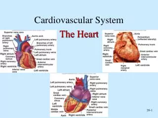
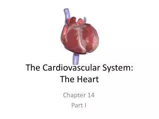
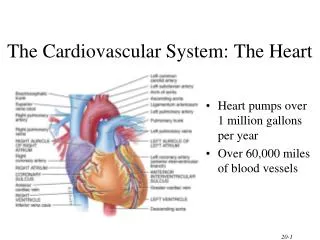
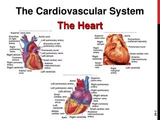
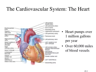
HEART LOCATION • Size, Location, and Orientation: • The heart is the size of a fist and weighs 250-300 grams • The heart is found in mediastinum and two-thirds lies left of the midsternal line • The base is directed toward the right shoulder and the apex points toward the left hip
Coverings of the Heart • The heart is enclosed in a double-walled sac called thepericardium: • The loosely fitting superficial part of this sac is the fibrous pericardium (tough, dense connective tissue) • Protects the heart • Anchors it to surrounding structures • Prevents overfilling of the heart with blood • Deep to fibrous pericardium is the serous pericardium: • Thin, slippery, two-layer serous membrane • The parietal pericardium lines the internal surface of the fibrous pericardium • Then parietal pericardium turns inferiorly and continues over the external heart surface as the visceral pericardium, or epicardium, which is an integral part of the heart wall • Between the parietal and visceral layers is the slitlike pericardial cavity, which contains a film of serous fluid • The serous membranes, lubricated by the fluid, glide smoothly past one another during heart activity, allowing the mobile heart to work in a relatively friction-free environment
HOMEOSTATIC IMBALANCE • Pericarditis: inflammation of the pericardium • Hinders production of serous fluid and roughens the serous membrane surface • Heart rubs against its pericardial sac creating a creaking noise (pericardial friction rub) that can be heard with a stethoscope • Deep pain to the sternum • Pericardia stick together and impede heart activity • Severe cases: • Large amounts of inflammatory fluid seeps into the pericardial cavity compressing the heart, limiting its ability to pump blood (cardiac tamponade) • Treated by inserting a syringe into the pericardial cavity and draining off the excess fluid
Layers of the Heart • The heart wall is composed of three layers, all richly supplied with blood vessels • Superficialepicardium is the visceral layer of the serous pericardium • Often infiltrated with fat, especially in older people • Middle layer, myocardium,is composed mainly of cardiac muscle and forms the bulk of the heart • It is the layer that contracts • Cardiac muscle cells are tethered to one another by crisscrossing connective tissue fibers and arranged in spiral or circular bundles reinforcing the myocardium internally and anchors the cardiac muscle fibers • The third layer, the endocardium (squamous epithelium), lines the chambers of the heart and is continuous with the endothelial linings of the blood vessels leaving and entering the heart
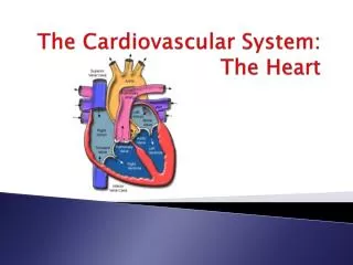
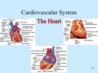
CHAMBERS and ASSOCIATED GREAT VESSELS • Heart has four chambers: • Two superior atria • Two inferior ventricles • Internal partition that divides the heart longitudinally is called the interatrial septum where it separates the atria, and the interventricular septum where it separates the ventricles • Right ventricle forms most of the anterior surface of the heart • Left ventricle dominates the infero-posterior aspect of the heart and forms the heart apex
CHAMBERS and ASSOCIATED GREAT VESSELS • Two grooves visible on the heart surface indicate the boundaries of its four chambers and carry the blood vessels supplying the myocardium • The atrioventricular groove, or coronary sulcus, encircles the junction of the atria and ventricles like a crown
CHAMBERS and ASSOCIATED GREAT VESSELS • The anterior interventricular sulcus, cradling the anterior interventricular artery, marks the anterior position of the septum separating the right and left ventricles • It continues as the posterior interventricular sulcus, which provides a similar landmark on the heart’s posteroinferior surface
Atria: The Receiving Chambers • Small, wrinkled, protruding appendages called auricles increase the atrial volume • Internally: • Posterior portion smooth-walled • Anterior portion the walls are ridged with bundles of muscle tissue (pectinate muscles) • Anterior and posterior regions are separated by a ridge called the crista terminalis • Interatrial septum bears a shallow depression (fossa ovalis), that marks the spot where an opening, the foramen ovale, existed in the fetal heart
Atria: The Receiving Chambers • Receiving chambers for blood returning to the heart from the circulation • Small, thin-walled chambers which contract only minimally to push blood “next door” into the ventricles • Blood enters the right atrium via three veins: • Superior vena cava returns blood from body regions superior to the diaphragm • Inferior vena cava returns blood from body areas below the diaphragm • Coronary sinus collects blood draining from myocardium • Blood enters the left atrium via four veins: • Pulmonary veins transport blood from the lungs back to the heart
Ventricles: The Discharging Chambers • Together the ventricles make up most of the volume of the heart • Marking the internal walls of the ventricular chambers are irregular ridges of muscle called trabeculae carneae which add support and strength • Papillary muscles play a role in valve function • Discharging chambers • Pumps of the heart • When ventricles contract, blood is propelled out of the heart into circulation: • The right ventricle pumps blood into the pulmonary trunk, which routes the blood to the lungs where gas exchange occurs • The left ventricle pumps blood into the aorta, the largest artery in the body, to the systemic trunk
Pathway of Blood Through the Heart • The right side of the heart pumps blood into the pulmonary circuit: • Blood returning from the body is relatively oxygen-poor and carbon dioxide-rich • Blood enters the right atrium and passes into the right ventricle, which pumps it to the lungs via the pulmonary arteries (conduct blood away from the heart) • In the lungs, the blood unloads carbon dioxide and picks up oxygen (oxygenated) • The left side of the heart pumps blood into the systemic circuit
Pathway of Blood Through the Heart • Freshly oxygenated blood from the lungs is carried by the pulmonary veins (toward the heart) back to the left side of the heart • Left side of the heart is the systemic circuit • Freshly oxygenated blood leaving the lungs is returned to the left atrium and passes into the left ventricle, which pumps it into the aorta • The aorta transports blood via smaller arteries to the body tissues, where gases and nutrients are exchanged across the capillary walls • Then the blood, once again loaded with carbon dioxide and depleted of oxygen, returns through the systemic veins to the right atrium via the superior vena cava and inferior vena cava
Pathway of Blood Through the Heart • Although equal volumes of blood are pumped to the pulmonary and systemic circuits at any moment, the two ventricles have unequal work-loads: • Pulmonary circuit, served by the right ventricle, is a short, low-pressure circulation • Systemic circuit, associated with the left ventricle, takes a pathway through the entire body and encounters about five times as much friction, or resistance to blood flow
Pathway of Blood Through the Heart • Functional differences of the two ventricles are revealed in their anatomy • The walls of the left ventricle are three-four times as thick as those of the right ventricle, and its cavity is nearly circular • The right ventricular cavity is flattened into a crescent shape that partially encloses the left ventricle, much the way a hand might loosely grasp a clenched fist • Consequently, the left ventricle can generate much more pressure than the right and is a far more powerful pump
Coronary Circulation • The heart receives no nourishment from the blood as it passes through the chamber: • The myocardium is too thick to make diffusion a practical means of nutrient delivery • The coronary circulation provides the blood supply for the heart cells: • The arterial supply of the coronary circulation is provided by the right and left coronary arteries, both arising from the base of the aorta and encircling the heart in the atrioventricular groove
Coronary Circulation • The left coronary artery runs toward the left side of the heart and then divides into its major branches: • Anterior interventricular artery : follows the anterior interventricular sulcus and supplies blood to the interventricular septum and anterior walls of both ventricles • Circumflex artery: supplies the left atrium and the posterior walls of the left ventricle
Coronary Circulation • The right coronary artery: courses to the right side of the heart, where it also divides into two branches • Marginal artery: serves the myocardium of the lateral right side of the heart • Posterior interventricular artery: runs to the heart apex and supplies the posterior ventricular walls • Near the apex of the heart, this artery merges (anastomoses) with the anterior interventricular artery • Together the branches of the right coronary artery supply the right atrium and nearly all the right ventricle
CORONARY CIRCULATION • The arterial supply of the heart varies considerably • Example: • 15% of people, the left coronary artery gives rise to both the anterior and posterior interventricular arteries • 4% of people, a single coronary artery supplies the whole heart • There may be both right and left marginal arteries • There are many anastomoses among the coronary arterial branches: • These fusing networks provide additional (collateral) routes for blood delivery to the heart • Explains how the heart can receive adequate nutrition even when one of its coronary arteries is almost entirely occluded • Even so, complete blockage of a coronary artery leads to tissue death and heart attack
CORONARY CIRCULATION • After passing through then capillary beds of the myocardium, the venous blood is collected by the cardiac veins, whose paths roughly follow those of the coronary arteries • These veins join together to form an enlarged vessel called the coronary sinus, which empties the blood into the right atrium • Obvious on the posterior aspect of the heart
CORONARY CIRCULATION • The sinus has three large tributaries: • Great cardiac vein: in the anterior interventricular sulcus • Middle cardiac vein: in the posterior interventricular sulcus • Small cardiac vein: running along the heart’s right inferior margin • Additionally, several anterior cardiac veins empty directly into the right atrium anteriorly
HOMEOSTATIC IMBALANCE • Blockage of the coronary arterial circulation can be serious and sometimes fatal • Angina pectoris: • Thoracic pain caused by a fleeting deficiency in blood delivery to the myocardium • May result from stress-induced spasms of the coronary arteries or from increased physical demands on the heart • Myocardial cells are weakened by the temporary lack of oxygen but do not die • Myocardial infarction (MI): • There is prolonged coronary blockage that leads to cell death • Commonly called a heart attack or coronary • Because adult cardiac muscle is essentially amitotic, most areas of cell death are repaired with noncontractile scar tissue • Whether or not a person survives a myocardial infraction depends on the extent and location of the damage • Damage to the left ventricle, which is the systemic pump, is most serious
Heart Valves • Blood flows through the heart in one direction: from atria to ventricles and out the great arteries leaving the superior aspect of the heart • This one-way traffic is enforced by valves that open and close in response to differences in blood pressure on their two side
Heart Valves • Two atrioventricular (AV) valves, one located at each atrial-ventricular junction ( tricuspid and bicuspid valves) prevent backflow into the atria when the ventricles contract • Right AV valve (tricuspid) has three flexible cusps (flaps of endocardium reinforced by connective tissue cores) • Left AV valve (bicuspid) has two flexible cusps • Commonly called the mitral valve because of its resemblance to the two-sided bishop’s miter or hat
Heart Valves • Attached to each AV valve flap are tiny white collagen cords called chordae tendineae (heart strings): • Anchor the cusps to the papillary muscles protruding from the ventricular walls
Heart Valves • Blood returning to the heart fills atria, putting pressure against AV valve • AV valve opens • When the heart is relaxed, the AV valves are open hanging limply into the ventricular chambers below and blood flows into the atria and then through the open AV valves into the ventricles • When ventricles contracts, compressing the blood in their chambers, the intraventricular pressure rises, forcing the blood superiorly against the valve flaps • As a result, the flaps edges meet, closing the AV valve
Heart Valves • The chordae tendineae and the papillary muscles serve as guy-wires to anchor the valve flaps in their closed position • If the cusps were not anchored in this manner, they would be blown upward into the atria, in the same way an umbrella is blown inside out by a gusty wind