Download
1 / 18
180 likes | 370 Views
Focused ultrasound reduces epileptic EEG bursts. Department of Radiology Brigham and Women's Hospital Harvard Medical School Byoung-Kyong Min. Introduction Method Result Discussion. Treatment on Neurological Disorders.

E N D
Focused ultrasound reduces epileptic EEG bursts Department of Radiology Brigham and Women's Hospital Harvard Medical School Byoung-Kyong Min
Introduction Method Result Discussion Treatment on Neurological Disorders • Representative non-invasive treatmenton the brain: Medication • However, it has side-effect and non-spatially specificity
Introduction Method Result Discussion Non-pharmacological neuro-modulation • Invasive tools (e.g. EpCS, DBS) • Non-invasive tools (e.g. TMS, tDCS) EpCS DBS Adapted from Hoy and Fitzgerald, Nature Review/Neurology, 2010 TMS tDCS Adapted from Hoy and Fitzgerald, Nature Review/Neurology, 2010
Introduction Method Result Discussion L1 L2 Focused-Ultrasound Sonication (FUS) • Image-guided, non-invasive, spatially-accurate focused-ultrasound (FUS) could be a potent tool for neuro-modulation. IR Marker Transducer Laser guide Motion camera Exablate (Insightec and GE): Array of small 1000 transducers
Introduction Method Result Discussion Idea: Pulsed application of FUS Previous observations by FUS • FUS suppresses VEP in LGN-sonicated cats (Fry, et al. 1958) • FUS affects the neurophysiology of in vitrolocal neural circuitry (Bachtold, et al. 1998, Rinaldi, et al. 1991) • FUS can temporarily modify the excitability of the neuronal tissue (Gavrilov et al. 1996) To avoid heating the tissue, pulsed sonication is used rather than continuous sonication. Fig. TBD: tone-burst-duration, PRF: pulse repetition frequency, AI: acoustic intensity
Introduction Method Result Discussion FUS transducer Superior A Inferior B B Rostral * Caudal C A Right Right Left Left Minutes Pre Sonication Post Sonication Recovery A B 1. Excitation (Yoo et al., 2008; 2009) Fig. FUS-mediated fMRI activation maps of the motor cortex (A & B) and FUS-mediated BOLD signal time course (C: gray bar: sonication) 2. Suppression (Yoo et al., 2008; 2009) Fig. Visual evoked potentials (A) and normalized amplitudes of the p30 components (B)
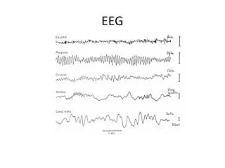
Introduction Method Result Discussion Application to Epilepsy suppression • We were motivated to examine if the FUS could suppress hyper-excitability of neural tissue based on a chemical kindling model of acute-stage epilepsy. • Epilepsyis a chronic neurological disorder (~50 million), and is characterized by seizures (abnormal hyper-excitability of neurons). • Since PTZ (pentylenetetrazol) was used to induce epileptic activity and progressive increments of theta activity has been reported during PTZ-induced epilepsy, not only raw EEG but also its theta band was assessed.
Introduction Method Result Discussion Experimental Setup & Design • Sprague–Dawley rats (275±30g) • Group 1 (PTZ(+)/FUS(+); n=9), Group 2 (PTZ(+)/FUS(-) ; n=9), Group 3 (PTZ(-)/FUS(+) ; n=9) • PTZ :GABAA receptor antagonist 45 mg/kg in 0.4 mL saline Fig. A diagram of the experimental apparatus
Introduction Method Result Discussion Transducer Characterization Transducer Hydrophone Fig. Transducer characterization • Mechanical Index (MI): the maximum peak negative pressure (Pr,α) of an ultrasound longitudinal wave divided by the square root of its center frequency (CMI) • FUS transducer: 690 KHz, 7cm ROC, 6cm OD, 0.5 ms TBD, 100Hz PRF, 130 mW/cm2 (Ispta)=2.6 W/cm2 (Isppa)
Introduction Method Result Discussion • EEG measures: sub-dermal electrodes (5 mm lateral to the midline & 7 mm anterior to the lambda), 1KHz sampling rate • Counting the number of raw EEG and theta bursts (4-8 Hz) exceeding the determined threshold (baseline σ × 4.75) in each session. • Racine scoring & Histological analysis • Statistics: Independent t-test (one-tailed) between the two groups, and paired t-test (one-tailed) within each animal. In order to compare body weights, a repeated-measures ANOVA with a covariance of the individual body weight before the experiment was applied. Anesthesia Full ictal PTZ Block-F Block-B Block-C Block-D Block-E Block-A Fig. Flowchart of the EEG acquisition and FUS sonication Baseline (10 min) Pre-FUS (10 min) Post1 (10 min) Post2 (10 min) FUS1 (3min) FUS2 (3min)
Introduction Method Result Discussion A. Sample EEGs from Group 1 (PTZ(+)/FUS(+)) 100 μV 10 sec FUS1 FUS2 Raw EEG 100 μV 20 μV EEG theta 1 min 20 μV 10 sec Fig. The sample time-courses of EEG recordings from PTZ-induced epileptic rats with sonication.
Introduction Method Result Discussion B. Sample EEGs from Group 2 (PTZ(+)/FUS(-)) 100 μV 10 sec 100 μV Raw EEG 20 μV EEG theta 1 min 20 μV 10 sec Fig. The sample time-courses of EEG recordings from PTZ-induced epileptic rats without sonication.
Introduction Method Result Discussion A Group Analysis • After sonication, the number of epileptic EEG bursts decreased. (‘Post1’: t(16)= -1.74; ‘FUS2’: t(16)= -2.03;‘Post2’: t(16)= -1.72). • After 2nd sonication, the number of theta EEG bursts decreased. (‘Post2’: t(16)= -1.98) B Fig.Comparison of the average number of threshold-exceeding raw (upper) and theta (lower) EEG peaks between the FUS-treated and untreated groups.
Introduction Method Result Discussion Summary • The number of epileptic EEG bursts within the FUS-treated group was significantly reduced after the sonication period (‘Post1’: t(8)= 2.26; ‘FUS2’: t(8)= 1.91; ‘Post2’: t(8)= 2.58). • The number of EEG theta peaks was significantly reduced during (63.0% reduction) and after (up to 68.5% reduction) the second sonication (‘FUS2’: t(8)= 2.81; ‘Post2’: t(8)= 3.14). • Racine scores of the FUS-treated group during a day after the experiment were significantly lower than those of the control group (t(15)= -2.41; FUS-treated group: 0.33; Control group: 1.13).
Introduction Method Result Discussion Histological Analysis Fig. Exemplary histological data obtained from Group 3 (left) H&E staining (right) TUNEL staining (DAPI in blue, apoptotic cell in green)
Introduction Method Result Discussion Discussion • The low-power, pulsed FUS sonication suppressed the number of epileptic EEG signal bursts without any significant tissue damages. • Stretch-sensitive ion channels (e.g. the novel chloride channels) may be involved in modifying the excitability of neural tissue. • Local hyperpolarization of the cell membrane would eventually raise the threshold for eliciting the epileptogenic activity. • Synaptic contacts could be disrupted by ultrasound, reducing the propagation of the epileptic activity across the brain. • Regulation of thalamic GABAergic inhibitory interneurons; PTZ a GABAA receptor antagonist • Therefore, FUS could provide a new non-invasive treatment of epileptic seizure.
Introduction Method Result Discussion Future Works • Intra-brain injection of KA (e.g. Hippocampus or amygdala) and evaluation of FUS on suppression of chronic focal epilepsy. • Assessment of neurotransmitter modulation associated with sonication (Microdialysis). • Stereotactic guidance: MRI-compatible stereotactic positioning system Acoustic radiation force impulse (ARFI) imaging
Introduction Method Result Discussion Acknowledgements • Seung-Schik Yoo, Krisztina Fischer, Yongzhi Zhang, Ferenc A. Jolesz: Department of Radiology, Brigham and Women’s Hospital, Harvard Medical School, Boston, MA, USA • Alexander Bystritsky: The Semel Institute for Neuroscience and Human Behavior, UCLA, LA, CA, USA • Kwang-Ik Jung: Department of Physical Medicine & Rehabilitation, Hallym University Sacred Heart Hospital, Korea • Lee-So Maeng, Sang In Park, Yong-An Chung: Institute of Catholic Integrative Medicine (ICIM), Incheon Saint Mary’s Hospital, Korea