Download

1 / 57
610 likes | 1.2k Views
Urine Analysis. Urine. Urine is formed in the kidneys, is a product of ultrafiltration of plasma by the renal glomeruli. Collection of Urine. Early morning sample-qualitative Random sample- routine Midstream sample-UTI Post prandial sample-D.M

E N D
Urine • Urine is formed in the kidneys, is a product of ultrafiltration of plasma by the renal glomeruli.
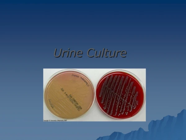
Collection of Urine • Early morning sample-qualitative • Random sample- routine • Midstream sample-UTI • Post prandial sample-D.M • 24hrs sample-preservatives like Toulene, thymol, chloroform or 2N HCl, Formalin are used. HCl- suitable for estimation of urea, ammonia and calcium thymol- for estimation of sodium, potassium, chloride, urea, protein, amylase and reducing sugars. • For quantitative estimation of proteins • For estimation of vanillylmandelic acid, 5-hydroxyindole acetic acid, metanephrines • For detection of AFB in urine • For detection of microalbuminuria
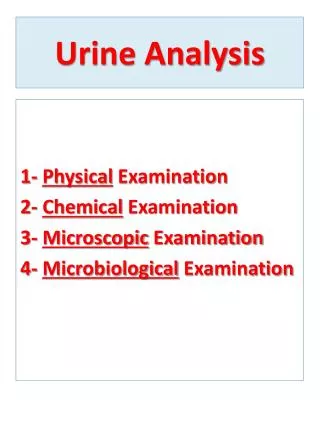
Urine examination • Macroscopic examination • Chemical examination • Microscopic examination
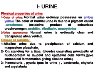
Macroscopic examination • Volume • Color • Odor • Reaction or urinary pH • Specific gravity
Volume of Urine • Normal =800-2,500 ml/day with an average of 1500 ml/day. • Polyuria- >2500ml • Oliguria-<500ml • Anuria-complete cessation of urine(<200ml) • Nocturia-excretion of urine by a adult of >500ml with a specific gravity of <1.018 at night (characteristic of chronic glomerulonephritis) Causes of polyuria • Diabetes mellitus and diabetes insipidus • Polycystic kidney disease, Chronic renal failure • Diuretics, Intravenous saline/glucose Causes of oliguria • Dehydration-vomiting, diarrhoea, excessive sweating • Renal ischemia, Acute renal failure • Acute tubular necrosis • Obstruction to the urinary tract
APPEARANCE NORMAL URINE- CLEAR • Cloudy- • Precipitation of amorphous phosphates in alkaline urine / amorphous urates in acid urine. • Amorphous phosphates dissolve on addition of acetic acid. • Amorphous urates will dissolve when specimen is heated. • Turbid- Leucocytes , epithelial cells, bacteria • Hazy- Mucous • Smoky- RBC • Milky- Fat, Chyle
Urinary pH/ Reaction • Reaction reflects ability of kidney to maintain normal hydrogen ion concentration in plasma & ECF • Normal= 4.5-8 with a mean of 6.0 in 24 hours • Tested by- 1.litmus paper 2. pH paper 3. dipsticks Acidic urine: • Ketosis-diabetes, starvation, fever • Systemic acidosis • UTI- E.coli • Acidification therapy • Alkaline urine • Strict vegetarian • Systemic alkalosis • UTI- Proteus • Alkalization therapy
Odor • Normal= aromatic due to the volatile fatty acids • AmmoniacalOdor- On keeping sample for a long time- bacterial action • Acetone like Odor- Ketonuria such as Diabetic ketoacidosis or starvation • Foul smell due to bacterial infections • Normal-1.016-1.025 • Depends on the concentration of various solutes in the urine. • Measured by-urinometer - refractometer - dipsticks Specific Gravity
Highspecific gravity 1.025 or more • All causes of oliguria Low specific gravity(hyposthenuria) 1.016 or less All causes of polyuria except glycosuria Isosthenuria=1.010 • Neither greater (more concentrated) nor less (more dilute) than that of protein-free plasma, typically 1.008-1.012. • Seen in chronic renal disease when kidney has lost the ability to concentrate or dilute urine (renal tubular damage/failure of renal medullary function)
Constituents of urine • Urine contains large number of inorganic and organic substances which are called normal constituents. • In a diseased state or pathological condition some substances are present in addition which are called abnormal constituents. • Normal constituents of urine: Inorganicconstituents: Sodium: 3-5g/day Chloride:10-15g/day Potassium:2-2.5g/day Calcium:0.1-0.3g/day Phosphates:0.8-1.3g/day Sulphates:1.0-1.2g/day Ammonia: 0.7-0.8g/day Organic constituents: Urea: 25-30g/day Uric acid: 0.5-0.8 g/day Creatinine: 1-1.8g/day Hippuric acid-0.7-0.8g/day
Abnormal constituents of urine • In a diseased state or pathological condition abnormal constituents are seen in urine. For example, in jaundice bilirubin is found in urine. In diabetes mellitus, glucose is excreted through urine. During nephritis, albumin is present in urine. • Different abnormal constituents of urine are: • Sugars • Proteins • Ketone bodies • Bile pigments • Bile salts • Blood
URINE GLUSOSE QUALITATIVE ESTIMATION: • Benedict’s test • Fehling’s test QUANTITATIVE ESTIMATION: • Benedict’s quantitative reagent method (BQR method)
BENEDICT’S TEST: • Principle-Benedict’s reagent contains CuSO4. Alkaline medium is provided to the reaction by sodium carbonate present in reagent . In the presence of reducing sugars cupric ions are converted to cuprous oxide which is hastened by heating, to give the color. • Method- take 5ml of Benedict’s reagent in a test tube, add 8drops of urine. Boil the mixture. • Result- blue-sugar absent; green-0.5% sugar; yellow-1% sugar; orange-1.5% sugar; brick red-2 % or more sugar.
Fehling’s test: Principle- • CuSO4 + 2NaOH Cu(OH)2 + Na2SO4 Blue colour • Cu(OH)2 + glucose 2CuOH + H2O + gluconic acid Yellow colour • 2CuOH Cu2O + H2O red colour During the reduction process of Cu(OH)2 to CuOH by the glucose. CuOH and its degradation product, Cu2O, are coloured compounds. Method- 2ml of fehling’s A + 2ml fehling’s B + 2-3 ml urine boil for 2-5 minutes
Benedicts quantitative reagent method (BQR method) • Benedict’s quantitative reagent composition: Sodium/Potassium citrate 200 gm+ Copper sulphate 18gm + Sodium carbonate100-200 gm+ Potassium thiocyanate + Potassium ferrocyanide. • Principle: BQR contains copper sulphate, potassium thiocyanate and other chemicals in alkaline solution. Copper ions of BQR are reduced to cuprous oxide by reducing monosaccharide glucose i.e., glucose in presence of Na2CO3 undergoes tautomerisation and forms powerful reducing agents enediols. These enediols show their reducing actions and reduces cupric ions to cuprous oxide and they get oxidised into gluconic acid. • Cuprous oxide is maintained in solution by potassium ferrocyanide and it reacts with potassium thiocyanate and forms a white ppt of cuprous thiocyanate instead of usual red ppt of cuprous oxide.
Disappearance of blue color from solution indicate complete reduction of copper sulphate and end point of the titration. 2Cu(OH)2 Cu2O+ H2O reduction Cu2O+ KSCN CuSCN (cuprous thiocyanate) -white ppt Method: • Pipette out 2ml BQR into 100ml conical flask, add 1ml water and 1 gm of anhydrous sodium carbonate mix well and add few pieces of porcelain. Flask is heated and heating is continued throughout titration. • Fill the burette with diluted urine sample. • Titrate the contents of the flask with the given sample, and note down the titer value. • Do the titration for 3 to 4 times to get concurrent values. • End point is blue to colorless. • The volume of urine used for complete titration is noted.
CLINICAL SIGNIFICANCE Presence of glucose in urine is known as glucosuria. Persistent glucosuria indicates the presence of diabetes mellitus. Normal value of glucose excretion in urine is 78.5mg/day In general glucose is seen in urine in 2 conditions A. When blood sugar is elevated B. When blood sugar is not elevated but renal tubular absorption-glucose is impaired. It is increased in • Any cause of increased blood glucose. • Rapid intestinal absorption (post gastrectomy dumping, normal pregnancy) • Endocrine disorders other than diabetes milletus like thyrotoxicosis, acromegaly, Cushing syndrome. • Major trauma, stroke, myocardial infarction or circulatory collapse, cerebral hemorrhage • Burns, oral steroid therapy, infection, pheochromocytoma. • Glycogen storage disease, obesity, sepsis, carcinoma of pancreas, fanconi’s syndrome, cystinosis.
Urine proteins • Urine normally contains only a scant amount of protein which derives both from blood and urinary tract itself. • Mainly albumin is filtered from nephrons due to low molecular weight, others are reabsorbed by renal tubules. • Other protein includes serum or plasma globulin, mucus or mucin, hemoglobin, Bencejones protein (immunoglobulin light chains produced by neoplastic plasma cells). Qualitative tests: • Sulphosalicyclic acid test • Heller’s nitric acid ring test • Heat coagulation test and acetic acid test Quantitative test for albumin: • Esbach’s method
Qualitative tests Sulphosalicyaclic Acid Test: Principle: It lowers the pH of the medium of the protein, so that both globular proteins become cationic and will react with an ionic sulphosalicylic acid and forms precipitate. Method: 3-4ml of clean urine + From the side of tube add 2-3 drops of 3%sulphosalicylic acid. Let it stand for 5 minutes. Observe the turbidity. Result: Formation of turbidity indicates presence of protein. Trace cloudiness against dark background • 1+ dense cloudiness. • 2+ cloudiness with granules and definite flocculation. • 3+ cloudiness with flocculation. • 4+ cloudiness with precipitation.
Heller’s test: 3ml of conc HNO3 and add urine from sides of the test tube- white ring at the junction of two fluids Principle: When protein solution is treated with strong acids like conc.HNO3, H2SO4 and HCl forms precipitate possibly due to change in pH or denaturation of protein. The denatured protein is less soluble in water and gets precipitated Heat & Acetic Acid Test: • Principle-proteins are denatured & coagulated on heating to give white cloud precipitate. • Method-take 2/3 of test tube with urine, heat only the upper part keeping lower part as control. • Presence of phosphates, carbonates, proteins gives a white cloud formation. Add acetic acid 1-2 drops, if the cloud persists it indicates it is protein(acetic acid dissolves the carbonates/ phosphates)
Quantitative Estimation-Esbach’s method • Esbach’s reagent: 5g of picric acid and 10g of citric acid in 500ml water. • Principle: Precipitation of protein by picric acid. Procedure- • Fill Esbach’salbuminometerwith acidic urine upto mark U and reagent is added uptomark R. Stopper the tube. • Tube is shaken well by inversion. • Keep in standing erect position for 18-24 hours for the precipitate to settle down. • Reading of the length of ppt is taken indicated by markings present over the tube. • Albumin is expressed in gm/L of urine.
Clinical Significance Interpretation- Insignificant amounts of proteins are excreted in urine in normal health not exceeding 20-80 mg/dl. This small amount is not detectable by routine methods. • Under certain conditions, as much as 20 G or more proteins may be excreted per day in urine. • The most common type of proteinuria is albuminuria; hence proteinuria and albuminuria are used synonymously. When proteins appear in urine in detectable amounts, it is called proteinuria. It can be caused by- a) Increased glomerular permeability b) Reduced tubular reabsorption c) Increased secretion of proteins d) Increased concentration of low molecular weight proteins in the plasma
Proteinuria • Accidental Proteinuria: Due to contamination of urine with vaginal seminal discharge after prostatic massage and derivation from diseased condition of genital tract or bladder accidental proteinuria is seen. • Functional Proteinuria: Non-pathological proteinuria also called physiological albuminuria mainly seen in strenuous exercise, exposure to cold, Pregnancy, Alimentary, if person stand in upright position for longer period. (Postural or orthostatic proteinuria) C. Pathological Proteinuria: I. Pre Renal: Severe dehydration, Heart diseases, Ascites (due to increased intra-abdominal pressure), Severe anemia, Fever, Collagen diseases, Toxemia of pregnancy
II. Renal: All inflammatory, degenerative or destructive diseases of kidney; common ones are: Nephrotic syndrome, Pyelonephritis, Acute and Chronic glomerulonephritis , Nephrosclerosis, Tuberculosis of kidney, Renal failure. III. Post Renal – Also called false proteinuria because in these conditions proteins do not pass through the kidneys. Causes include- • Severe urinary tract infections • Inflammatory, degenerative or traumatic lesions of pelvis, ureters, bladder, prostate or urethra • Bleeding genito urinary tract • Pus in urine • Contamination of urine by semen or vaginal secretions
Microalbuminuria • Urinary albumin excretion between 30-300 mg/day. • Cannot be detected by dipstick methods. • Strong predictor of development of diabetic nephropathy. • Can be detected 10-15 years before development of diabetic nephropathy. • Significant risk marker of cardiovascular ds. • Measured by nephelometry and radioimmunoassay Diagnostic relevance microalbuminuria: • In diabetic patients for early diagnosis of nephropathy. • In hypertensive patients as indicator of end organ damage
Ketone bodies • The term ketones refer to 3 intermediate product of fat metabolism, they are acetone, acetoacetic acid and beta-hydroxybutyricacid. Acetone: Constitutes 2%. It is volatile & excreted primarily through the lungs DiaceticAcid (Acetoacetic): The first formed. • Both acetone and beta hydroxybutyric acid are produced from diacetic acid. • Diaceticacid is the form detected by most ketone test procedures. • Makes up 20 % of total.
Beta-hydroxybutyricAcid: • It is majorly formed (78%) • Although most of the ketones are this form, it is not detected by routine test. • Only Hart’s test, an old ‘wet chemical’ test that is designed to detect B-hydroxybutyricacid Ketone is found when there is excessive fat metabolism which occurs in various situations: • Impaired ability to metabolize carbohydrate • Inadequate carbohydrate intake • Excessive carbohydrate loss • Increased metabolic demand.
Methods: 1. Rothera's test for acetone. 2. Gerhardt's test for acetoacetic acid 3. Hart’s test for betahydroxybutyricacid. 1) Rothera’sTest Principle: Nitroprusside in alkaline medium reacts with a ketone group to form a purple ring. It is given by acetone and acetoacetate, but not by Beta-hydroxybutyric acid. Procedure: • Saturate 5 ml of urine with solid ammonium sulphateand add 0.5 ml of freshly prepared sodium nitroprusside (5%). • Mix well and add liquor ammonia from the side of tube. • A purple ring at the junction of the liquid indicates the presence of ketone bodies.
2) Gerhardt’s ferric chloride test Principle: A purplish color is given by acetoacetate. On boiling acetoacetate is converted to acetone and does not give this test positive. This test is only given by acetoacetate and not by beta- hydroxybutyrate. Procedure- • Add 10% ferric chloride solution drop by drop to 5 ml of urine in a test tube. • If phosphates are present, precipitates of ferric phosphates may form, that should be filtered off and the ferric chloride is added. • False positive Gerhardt’s test may be obtained with Salicylic acid and Salicylates.
3) Hart’s test for β- OH butyric acid This basically involves the conversion of beta- hydroxybutyric acid to acetone, which can then be detected by the nitro-prussidemethod. • First, 20 mL of urine is acidified with diluted acetic acid and then boiled until it becomes half of the original volume. This amount is cooled and then raised to the original volume with water. The purpose of this process is to remove the acetoacetic acid and acetone. • The specimen is then divided into two portions and put into two test tubes. • Next, 1 mL of hydrogen peroxide is added to the first portion, which is warmed gently and cooled. • In this process, beta-hydroxybutyric acid is changed to acetone. • Then ten drops of nitroprusside solution are added to both tubes and overlaid with ammonia. • The presence of beta-hydroxybutyric acid is indicated by a purple-red color reaction that occurs in the sample treated with hydrogen peroxide.
Clinical Significance Ketonemiaand hence ketonuria occurs mostly in conditions of glucose deprivation. Nondiabetic Ketonuria: Often due to the increased catabolism of adipose tissue when there is limited intake of food- Severe Starvation / Fasting/ Anorexia (loss of appetite); High fat feeding; Heavy exercise; Severe carbohydrate restriction • Ketonuriais frequently seen in infants or children with acute febrile diseases or toxic states which produce vomiting or diarrhea. • Also found when there is vomiting due to general ill health, pregnancy, or anesthesia. • Administration of a ketogenic diet to treat seizures in children, glycogen storage disease (Gierke's) and, occasionally, exposure to cold or severe exercise. • Toxemia of pregnancy; Propanol poisoning;; Fever; Lactic acidosis; Salicyclatetoxicity.
Diabetic Ketonuria: Ketonuria in diabetics indicates ketosis (diabetic acidosis), the possibility of an impending coma. Note: • In diabetes mellitus impaired ability to metabolize carbohydrate takes place. As carbohydrate cannot be used to meet the body energy need, fats are burned which leads to the presence of ketones in the urine. • Acetoacetic acid oxidizes rapidly to form acetone therefore test must be carried out in fresh urine specimen. • Individuals receiving levodopa, paraldehyde, pyridium and phathalein compound may produce false positive result when tested for ketonuria. Presence of salicylates give false negative result. • When sugar is found in urine, the urine should be tested for ketone.
Test for Bile salts Hay’s Sulphur test Principle: Bile salts lower the surface tension allowing the sulphur powder to sink. Procedure: Sprinkle a little dry sulphur powder on the surface of fresh urine in a test tube taking distilled water as control. Sulphur powder sinks in the presence of bile salts. Result: • In the control, sulphur powder remains immiscible with the underlying liquid. In the positive test, the sulphur powder sinks to the bottom. • Interpretation: Bile salts and bile pigments are present in urine in obstructive jaundice. Control for comparison Positive test
Bile salts Primary bile acids: • Cholic acid and chenodeoxycholic acid (CDCA)- synthesized from cholesterol in the liver, conjugated with glycine or taurine, and secreted into the bile. Secondary bile acids: • Deoxycholate and lithocholate, are formed in the colon as bacterial metabolites of the primary bile acids. • Sodium taurocholate and sodium glycocholate are found in urine.
heme→ biliverdin → bilirubin transport to the liver (albumin) conjugation with glucuronate bilirubin diglucuronide secreted into the bile bacteria in the small intestine release bilirubin from diglucuronide and convert it to colourless urobilinogens a small fraction is reabsorbed and re-excreted through theliver into the bile a small fraction is excreted into the urine by the kidney most of them are oxidized to pigments and excreted in the faeces (urobilin, stercobilin)
Tests for bile pigments 1. Fouchet’s test: Principle: BaCl2 reacts with sulphate in urine to form barium sulphate. If bilirubin is present in urine, it adheres to precipitate and is detected by oxidation with FeCl3 in the presence of trichloro acetic acid to form biliverdin (Green) Procedure: • Take 5 ml of 10% BaCl2 to 10 ml of urine and filter. • Dry the filter paper and add a few drops of Fouchet's reagent (Prepared by adding 10 ml of 10% FeCl3 to 100 ml of 25% TCA). • A green color is obtained due to oxidation of bilirubin to biliverdin.
2. Gmelin’stest: Principle: Nitric acid oxidizes Bilirubin to Biliverdin giving different colors from green to violet. Procedure: To about 5 ml of concentrated HNO3 in a test tube, add an equal volume of urine carefully so that the two liquids do not mix. At the junction of two liquids various colored rings (Green, blue, red, violet etc.) will be formed. 3. Iodine test: Procedure : Dilute some tincture of iodine with one to two volumes of water and layer it carefully on to some urine in a test tube, a green ring at the junction of two fluids indicates the presence of Bilirubin. It is not a sensitive test, cannot detect small amount of bilirubin present in the given sample.
Clinical significance • Bilirubin in urine means increased amount of conjugated bilirubin because unconjugated bilirubin is water insoluble and is also bound to albumin, hence cannot cross the glomerular membrane. • Bilirubin passes from the blood to the liver, where it becomes water soluble (conjugated), and enters into the bile ducts. It then enters the intestine with the bile. Normally, there is no bilirubin in the urine. • However, bilirubin may appear in the urine in cases of hepatitis, in cirrhosis, and in other conditions where there is moderate to severe hepatocellular damage • The bilirubin test can be used to differentiate between hemolyticjaundice and obstructive jaundice. • In cases of obstructive jaundice(intra or extra hepatic) urinary bilirubin is present; in cases of hemolytic jaundice bilirubin is characteristically absent from urine. Hence, urinary bilirubin is a useful indicator of the early phases of chemical or viral injury to the liver.
Test for Urobilinogen Ehrlich’s test: Principle: The test for urobilinogen is based on the Ehrlich Aldehyde Reaction. P-dimethylaminobenzaldehydein an acid medium with a color enhancer reacts with urobilinogen to form a pink-red color. The optimum temperature for testing is 22° - 26°C. A freshly voided sample is best for optimal results. Procedure: • Take 5 ml of fresh urine in a test tube and add 5 ml of Ehrlich's reagent to it. • Wait for 10 minutes and add 10 ml of saturated sodium acetate solution. • A pinkish color indicates the presence of urobilinogen. • Porphobilinogenis also detected by Ehrlich's test. The color intensifies upon addition of sodium acetate if Porphobilinogen is there.
Clinical significance • Urobilinogen is found in urine in hepatic and prehepatic jaundice. • It is present in excessive amount in prehepatic jaundice and is completely absent in post hepatic jaundice. • An increased urobilinogen concentration in urine is a sensitive index of liver dysfunction or hemolytic disorders.
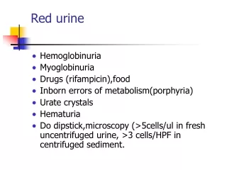
Blood in urine Presence of blood in urine is called hematuria- RBCs/haemoglobin. Haematuria- when 5 or more intact RBCs/HPF(high power field) a. Gross hematuria: Urine appears reddish in gross hematuria and this is observed in renal stones, malignancies, trauma, tuberculosis and acute glomerulonephritis. b. Microscopic hematuria: Blood is not visible to naked eyes. It is observed in: • Malignant hypertension, • Sickle cell anemia, • Coagulation disorders, • Polycystic kidney disease, • Incompatible blood transfusion, • Auto immune hemolytic anemia.
Causes of Haematuria Renal • Neoplasms • Calculi • TB • Pyelonephritis • Hydronephrosis • Oxaluria • Acute GN • Polycystic kidney disease • Post-Renal • Ureter- calculus, neoplasm • Urinary bladder- neoplasm, TB, Cystitis, calculus. • Prostate- BPH, Neoplasm • General • Embolism of kidney from SBE(Subacute bacterial endocarditis ). • Malignant HTN kidney • Haemophilia • Leukemia
Causes of Haemoglobinuria • Malaria- black water fever. • Hemolytic streptococcal septicaemia. • Incompatible blood transfusion. • Drugs- Sulphonamides, phenylhydralazine. • PNH (Paroxysmal nocturnal hemoglobinuria)
BenzidineTest Principle: The peroxidase activity of hemoglobin decomposes hydrogen peroxide releasing nascent oxygen which in turn oxidizes benzidine to give blue color. Reagents: Saturated solution of benzidine in glacial acetic acid, Hydrogen peroxide Procedure: • Add 2 ml of urine in test tube. • Add 2ml of 1% Benzidine solution in acetic acid. Shake well. • Add 2ml of hydrogen peroxide. • Mix and observe for a change in color. • Positive result: Green or blue color. (Hematuria)
DIPSTICK METHOD OF URINALYSIS • Seven different reagent areas are affixed on the strip. • These different cellulose areas are impregnated with specific testing chemicals according to the test which reacts with specific substances present in urine by changing the color. • The entire strip is dipped in the urine sample and color changes in each square are noted. The color change takes place after several seconds to a few minutes from dipping the strip. If read too early or too long after the strip is dipped, the results may not be accurate. • Color change chart is observed and compared to the color chart for the presence of abnormal levels of substances. • The various determinations done by this are pH, Specific Gravity, Glucose, Protein, Ketones, Urobilinogen, Blood, Bilirubin
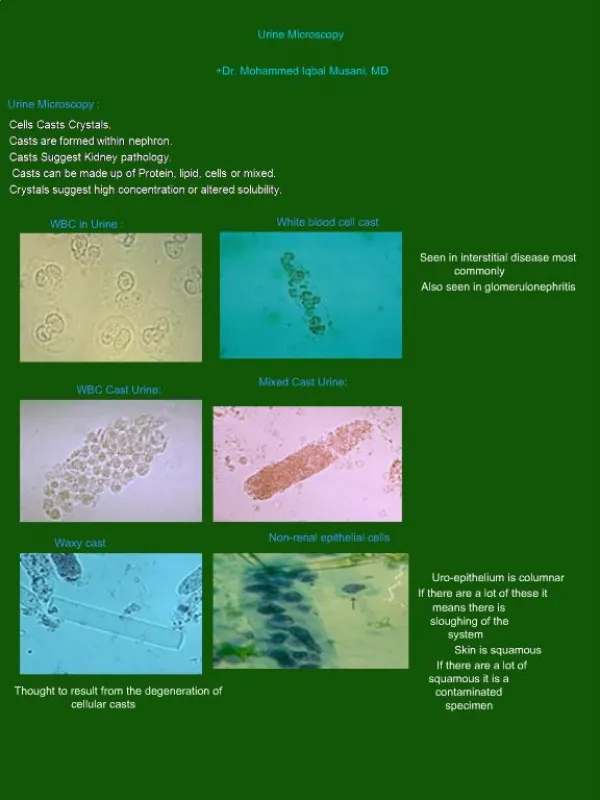
Microscopic analysis • Microscopic urinalysis is done by pouring the urine sample into a test tube and centrifuging it (spinning it down in a machine) for a few minutes. • The top liquid part (the supernatant) is discarded. • The solid part left in the bottom of the test tube (the urine sediment) is mixed with the remaining drop of urine in the test tube and one drop is analyzed under a microscope