Download

1 / 35
360 likes | 447 Views
This text explains embryonic development of the digestive system, covering folding stages, foregut, midgut, and hindgut formation. It details tissue origins, pharyngeal arches, and organ derivations, such as liver, pancreas, and intestines. Teratological conditions like atresia and stenosis are also discussed.

E N D
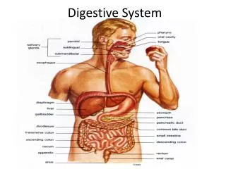
Embryo folding – incorporation of endoderm to form primitive gut. Outside of embryo – yolk sac and allantois. Vitelline duct
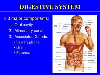
Stomodeum (primitive mouth) the oral cavity + the salivary glands Proctodeum primitive anal pit Primitive gut whole digestive tube + accessory glands Tracheo- esophageal septum Proctodeum
pharynx forgut midgut hindgut
Tissues in GIT • The epithelium of gut and glandular cells of associated glands of the gastrointestinal tract develop from endoderm • The connective tissue, muscle tissue and mesothelium derive from splanchnic mesoderm • The enteric nervous system develops from neural crest
primitive gut foregut midgut hindgut from above ductus to cloacal pharyngeal omphalomesentericus membrane membraneand yolk sack Pharyngeal membrane
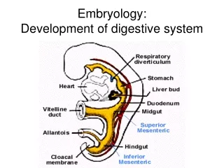
Derivatives of foregut – pharynx, (+ respiratory diverticle), esophagus stomach, cranial part of duodenum (+ liver, gall bladder pancreas), midgut – caudal part of duodenum, small intestine and part of large intestine (cca 1/3 of colon transv.) hindgut – the rest of large intestine, rectum, upper part of the anal canal
Liver bile duct pancreas Rectum Foregut Midgut Hindgut
Oral cavity • primitive mouth pit – stomodeum • lined with ectoderm • surrounded by: - processus frontalis (single)- proc. maxillares (paired)- proc. mandibulares (paired) • pharyngeal membrane(it ruptures during the 4th week, primitive gut communicates with amniotic cavity)
Pharyngeal (branchial) apparatus Pharyngeal arches • appear in weeks 4 - 5 • on the ventral side of the pharyngeal gut; • each arch contains cartilage, nerve, aortic arch artery and muscle; • pharyngeal clefts and pouches are located between the arches; • membrana obturans membrana obturans ectoderm endoderm
Fate of pharyngeal pouches and clefts Tympanic membrane + tympanic cavity Sinus cervicalis early later
Structures derived from Pouches Each pouch is lined with endoderm and generates specific structures.
Esophagus development below respiratory diverticle, behind larynx and trachea primitive pharynx thyroid gl. laryngotracheal diverticle (respiratory diverticle) esophagus
Esophagus development • differentiation of epithelium from endoderm • during the 8th week endoderm proliferates and temporarily closes esophageal lumen • other tissues and structures in the wall arise from splanchnic mesoderm
Mesenteries – suspensory duplicature derived from mesoderm and mesenchyme (a fold of tissue that attaches organs to the body wall) mesooesophageum dorsal wall of body esophagus mesoesophageum dorsale gives rise to dorsal mediastinum and mediastinal pleura mesoesophageum ventrale disappears
Teratology Esophageal atresia – failure of recanalization or septum deviation Susp.: polyhydramnios, fetus cannot swallow Esophageal stenosis – narrow lumen, incomplete recanalization Tracheoesophageal fistula – defect in septum
Stomach development 90º in the 4th week – spindle dilatation of distal forgut in median plane endoderm – epithelium and glandular cells splanchnic mesoderm – other tissues of stomach wall
Uneven growth of ventral and dorsal wall: - curvatura minor (to the right), - curvatura major (to the left). Rotation around sagital axis : - curvatura minor (cranial position), - curvatura major (caudal position). Rotation around longitudinal axis: - left side → ventrally, - right side → dorsally.
foregut midgut duodenum
Teratology Pyloric stenosis – muscular hypertrophy, unknown etiology Duodenal stenosis – incomplete recanalization Duodenal atresia – polyhydramnios vomiting
Midgut Thederivatives • thedistal duodenum, jejunum, and proximal ileum + • thedistal ileum, cecum, appendix, ascendingcolon, and proximal 2/3 oftransversecolon. themidgutgrowsfasterthanthe embryo, creating: • duodenalloop • umbilicalloop
Flexura duodenojejunalis Duodenal loop and umbilical loop forgut midgut Umbilical loop herniates into the umbilical cord (physiologic herniation, in week 6-10)
Duodenum development • Duodenal loop – 2 limbs: upper limb(from foregut) lower limb (from midgut) • Week 5 – 8, duodenum is temporarily obliterated • On top of loop – diverticles (for liver, gallbladder, pancreas)
Liver, gall bladder, pancreas The liver bud (hepatocystic diverticle) appears at the distal end of the foregut (week 4) and divides into hepatic and cystic diverticles, later ventral pancreatic bud and dorsal pancreatic bud (week 5). Both pancreatic buds meet and fuse (week 6). Septum transversum
Due to rotation of stomach, mesenteries and umbilical loop, duodenal loop changes its position (from front to the right) and becomes retroperitoneal organ (together with pancreas) Spleen - derives from mass of cells in dorsal mesogastrium during week 5
Intestines development • Umbilical loop – 2 limbs: cranial – jejunoileal limb (jejunum, major part of ileum) caudal – ileocecal limb (rest of ileum, caecum + appendix, colon ascendens and 2/3 of colon transversum) • A. mesenterica sup. – axis of rotation • week 6 – physiologic herniation into the umbilical cord, week 10 – reposition into abdominal cavity
90º after 270º rotation 180º • In theumbilicalcord, themidgutlooprotates90°counter-clockwisedirectionaroundthe axis ofthe superior mesentericartery. • Uponreturning, the gut undergoesanother180°counter-clockwiserotation, placingthececum and appendixneartheright lobe ofthe liver. • Thetotalrotationofthe gut is270°.
Hindgut The distal end of the hindgut – the cloaca. Derivatives of the hindgut: the distal 1/3 of the transverse colon, descending colon, sigmoid colon, rectum and upper part of anal canal (above the pectinate line).
Division of the cloaca - urorectal septum divides the cloaca into a ventral urogenital sinus and a dorsal anorectal canal. The cloacal membrane breaks down during the 7th week. Distal to the pectinate line (site of the former cloacal membrane), the epithelium of the anal canal derives from ectoderm of proctodeum (primitive anal pit)
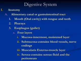
Mesenteries • double layer of peritoneum enclosing organs and connecting them to the body wall Ventral mesentery exists only in region of distal part of esophagus, stomach (lesser omentum) and upper part of duodenum Dorsal mesentery forms dorsal meso- gastrium (greater omentum), dorsal mesoduodenum, mesentery proper (jejunum, ileum)