Download
1 / 34
350 likes | 1.06k Views
Explore the detailed anatomy of the eye, including the layers, vessels, and functions of the eyeball's components like the fibrous layer, sclera, cornea, vascular layer, choroid, ciliary body, iris, and retina. Learn about the visual axes, optic axis, equator, and more.

E N D
THE EYEBALL Prof. Dr. Selda Önderoğlu Department of Anatomy
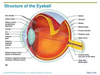
THE ORGAN OF VISION • Located in the orbit- embedded in orbital fat • Fascial sheath- capsule of Tenon • Two spheres - anterior segment --transparent , 1/6 - posterior segment -- opaque, 5/6
AXES OF EYEBALL OCULAR AXIS - anterior pole- posterior pole - parallel in two eyeballs OPTIC AXIS • posterior pole of lens- fovea centralis • Not parallel in two eyeballs EQUATOR OF EYEBALL
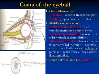
LAYERS (TUNICS) 1-FIBROUS LAYER 2-VASCULAR LAYER (uveal tract) 3-NERVOUS LAYER (retina)
FIBROUS LAYER- FUNCTIONS • A place for the attachment of muscles. • Maintain a constant intraocular pressure. • Protect the deeply located structures.
FIBROUS LAYER • SCLERA ( characteristics ) -5/6 POSTERIOR PART White part Opaque, hard Thickest behind . CORNEA -1/6 ANTERIOR PART Transparent
SCLERA • Tenon’s capsule (Fascial Sheath-Vagina Bulbi) • Episcleral space • SCLERA • Perichoroidal space- delicate cellular tiss. . VASCULAR LAYER
SCLERA • IS POSTERIORLY • PIERCED BY: • fibers of the optic nerve(LAMINA CRIBROSA • SCLERAE) • ciliary vessels and nerves • central vessels of retina • venae vorticosae ( 4-5 in number)
SCLERA meets the CORNEA at the • SCLEROCORNEAL JUNCTION ( LIMBUS CORNEA ) • SINUS VENOSUS SCLERAE ( SCHLEMM’S CANAL ) -At the sclerocorneal junction.
SCLERA Vessels ----- few. Nerves-------- ciliary nerves
CORNEA • The major site of refraction of light entering the eye. • 1/6 anterior part More convex. • Transparent. • Avascular. • Rich in nerves-opthalmic n. • Does not have lymph vessels.
VASCULAR LAYER(UVEAL TRACT ) • CHOROID • CILIARY BODY • IRIS
CHOROID • 5/6 posterior part of the eyeball. • highly vascular • Color– dark brown • Extends as far as the ora serrata. • Outer surface is related to—suprachoroid lamina • Inner surface—pigmented layer of retina
CILIARY BODY CHOROID continues anteriorly as the ciliary body
CILIARY BODY • Rich in blood supply • CILIARY PROCESSES secrete AQUEOUS HUMOUR • SUSPENSORY LIGAMENTS- towards the lens
CILIARY BODY-FUNCTIONS • SUSPENSION OF LENS • PRODUCTION OF AQUEOUS HUMOUR • CHANGING THE ANTERO-POSTERIOR DISTANCE OF LENS (FOR THE MECHANISM OF ACCOMODATION )
CILIARY MUSCLE • Smooth muscle- • Nerve-psymp- OCULOMOTOR- postganglionic fibers from ciliary ganglion • Function-changes in anteroposterior distance of lens. ---contract-susp. Lig. Relax.-thickness of lens increases. (or opposite)
IRIS • Coloured (blue- dark brown ) • Surrounds the pupil • Adjustable diaphragm • Has two margins - CILIARY - PUPILLARY
IRIS • Somewhat suspended in aqueous humour and divides the area between cornea and lens into two chambers • ---ANTERIOR ---POSTERIOR Cornea – iris meet at —IRIDOCORNEAL ANGLE.
AQUEOUS HUMOUR CILIARY PROCESSES -> POSTERIOR CHAMBER - > PUPIL –> ANTERIOR CHAMBER –> SINUS VENOSUS SCLERAE (at the iridocorneal angle) –> ANTERIOR CILIARY VEINS- > OPHTHALMIC VEINS –> CAVERNOUS SINUS
MUSCLES OF THE IRIS • SPHINCTER PUPILLA MUSCLE—smooth muscle-meiosis—innervated by parasymp.-short ciliary nn. ( oculomotor n.) • DILATOR PUPILLA MUSCLE—smooth muscle- midriasis- innervated by symp.—sup. cervical symp. gang.-int. car. a.-long ciliary nn.
ARTERIES OF IRIS • long posterior ciliary aa.-(ophthalmic a.) • Short posterior ciliary aa.-(muscular brs.) --MAJOR ARTERIAL CIRCLE—(at the ciliary margin) Brs. From major arterial circle and anterior ciliary aa. --MINOR ARTERIAL CIRCLE
RETINA • Innermost layer of the eyeball. • The neural- sensory layer • Composed of 2 layers ----outer—PIGMENTED LAYER ----inner---NERVOUS LAYER
ORA SERRATA • The place where nervous layer of retina ends. • But the pigmented layer continues anteriorly over the back of the ciliary processes and the iris—ciliary part-iridial part.
PARTS OF RETINA OPTIC PART from optic disc-to ora serrata pigmented+ nervous layers present here. PARS CAECA RETINA-Blind part. from ora serrata Anteriorly.Only pigmented layer present.
MACULA LUTEA-FOVEA CENTRALIS • MACULA LUTEA=YELLOW SPOT • Oval yellowish area near the the centre of posterior part of retina. • FOVEA CENTRALIS-is the central depression located in the macula lutea---where VISUAL RESOLUTION IS HIGH. HAS ONLY CONES.
OPTIC DISC-araea where optic n. Leaves the eyeball • OPTIC DISC = BLIND SPOT insensitive to light. 3mm. Nasal to macula lutea. --central a. And v.of retina. ---in normal ophthalmoscopic examinations color= PINK --- if optic n. Atrophies,color = WHITE
CENTRAL RETINAL ARTERY • Courses through optic nerve fibers • The first branch of ophthalmic artery • Branches – superior and inferior each divides into temporal & nasal brs. -So 4aa. Supplies each quadrant of retina.
CENTRAL RETINAL VESSELS • ARE THE ONLY VESSELS IN THE BODY WHICH CAN BE INSPECTED DIRECTLY WITH NAKED EYE ( through an ophthalmoscope).
OCULAR REFRACTIVE MEDIA • CORNEA • AQUEOUS HUMOUR • LENS • VITREOUS HUMOUR
AQUEOUS HUMOR • Fills the anterior and posterior chambers. • Secreted by ciliary processes- Functions -intraocular pressure -shape of eyeball -metabolic avenue for avascular structures- cornea, lens.
VITREOUS HUMOR • Occupies vitreous body (chamber)- posterior to lens. • Colorless,structureless,transparent gel
LENS • Biconvex, transparent body. • Has a capsule. • Central points - anterior pole - posterior pole-AXIS equator, faint sutural lines(radii lentis) devoid of vessels.
ARTERIES AND VEINS OF EYEBALL • Artery- Ophtalmic artery • Veins- Opthalmic veins