Download
1 / 1
20 likes | 190 Views
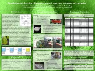
Inoculation and detection of Impatiens necrotic spot virus in tomato and cucumber. Kristin Vickers, Dr. Linda Hanley-Bowdoin, Mauricio Montero,and Natalia Barboza Vargas. 1 North Carolina State University, Raleigh, NC and 2 University of Costa Rica, San Jose, Costa Rica. Introduction.

E N D
Inoculation and detection of Impatiens necrotic spot virus in tomato and cucumber Kristin Vickers, Dr. Linda Hanley-Bowdoin, Mauricio Montero,and Natalia Barboza Vargas.1North Carolina State University, Raleigh, NC and 2University of Costa Rica, San Jose, Costa Rica. Introduction Results and Discussion Materials and Methods • Inoculation • 7 INSV samples to inoculate with: • T101, T107, T152, T154, T156 • China (C+), Buffer (C-) • Selected 42 plants to inoculate • 21 tomato • 21 cucumber • Ground tissue from samples with 3% NaSO3 with a mortar and pestle • Apply solution onto selected plants • Spray with carborundum • Rub final layer of liquid on plants • Tissue Collection • Collected samples • from each plant • Ground tissue into liquid to be placed in microtubes • ELISA • Day 1 • Make a solution of antibody • and carbonate buffer • Add 100µL to each well • Day 2 • Clean plates by emptying and filling with wash buffer • Fill with 100µL of sample Impatiens necrotic spot virus (INSV)is part of the genus Tospovirus and the family Bunyaviridae. This virus is one of the most devastating to many important vegetables, legumes, and ornamental plants throughout the world, particularly in Asia and South America (Mandal, 2012). Nineteen species of Tospovirus have been discovered up to this point. There were two positives as a result from the spectrophotometer, which were labled as T152t.b and T154p.b (Table 1). All of the species within the Tospovirus genus are transmitted by ten different species of thrips through a circulative and propagative manner. These viruses must be presented to the first or second larval stages for the adult thrip to emerge and transmit the virus, providing a “dead-end” epidemiology. Visual symptoms include necrosis, spotting, rings forming, lesions, yellow pale streaks, and reduction in yield and plant size (Pappu, 2009). Diagnosing this virus can be done using a common serological detection test, enzyme-linked immunosorbent assay (ELISA). The Sandwich ELISA virus detection test uses an antibody/enzyme combination to trap the specific antigens of a specific virus with color change to determine if positive for that virus (Clark, 1977). Table 1. Final ELISA results for tomato and cucumber. T Future Work The low positives could have been caused by inoculation, frozen samples used, rejection by defense system, or time constraint.I would like to have performed the ELISA process again after one more week with hopes of higher number of positive results. Objectives Acknowledgements The overall goals of this project were : To inoculate economically or experimentally important plant species with a set of INSV isolates for their biological characterization. To confirm mechanical inoculation results by means of ELISA (enzyme-linked immunosorbent assay). Get to know the techniques for data analysis and presentation. This project was funded by the National Science Foundation, as part of the IRES (International Research Experiences for Students) program. • Day 3 • Clean with wash buffer • Add conjugate antibody to wells • Incubate • Substrate is added • Run plate in spectrophotometer ** Presented at the: 22nd Annual Undergraduate Research Symposium, North Carolina State University, July 2013, Raleigh, NC.