Stability Assessment of Chitosan Nanoparticles in Physiologically Relevant Storage Solutions
This study investigates the stability of chitosan nanoparticles, prepared via pH-controlled precipitation and ionic crosslinking with sodium tripolyphosphate (TPP). Utilizing dynamic light scattering (DLS) for analysis, nanoparticles were stored in various environments: distilled water, PBS at pH 6.8, PBS at pH 7.4, and 10% FBS-DMEM. Results indicate that TPP-crosslinked chitosan nanoparticles exhibit superior stability in relevant physiological conditions compared to those prepared by pH-controlled methods. These findings underscore the importance of stability considerations for biomedical applications.

Stability Assessment of Chitosan Nanoparticles in Physiologically Relevant Storage Solutions
E N D
Presentation Transcript
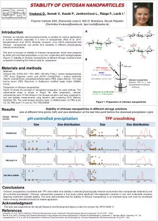
A Chitosan, as naturally derived polysaccharide, is suitable for various applications in human medicine, especially in a form of nanoparticles(Riva et al. 2011; Sanyakomdhornet al. 2013). Recently, Jonassen et al. (2012) emphasized that chitosan nanoparticles can exhibit time instability in different physiologically relevant environments. This work is focused on stability of chitosannanoparticles, which were prepared by either pH-controlled precipitation or by ionic crosslinking with tripolyphosphate (Figure 1).Stability of chitosan nanoparticles at different storage conditions were compared considering the method used for preparation. rate of addition = 0.2 mL / 1 min B Materials Chitosan (No. 50494, DD = 76%, MW = 450 kDa, Fluka), sodium tripolyphosphate (TPP, Acros Organics), acetic acid (AcOH, CentralCHem ) sodium hydroxide (NaOH,CentralCHem), phosphate-buffered saline (PBS, Sigma Aldrich), 10% fetal bovine serum (FBS, Biochrom) in Dulbecco’s modified eagle media (DMEM,Lonza). Chitosannanoparticles Chitosan : TPP = 9 : 1 w/w Concentration of chitosan 0.5% w/v Preparation of chitosan nanoparticles Figure 1A shows the principle of nanoparticle preparation by used methods. The experimental setup is shown in Figure 1B. After preparation, chitosan nanoparticles were 10-fold diluted in the storage solutions and were subjected to size and stability measurements using dynamic light scattering (DLS, Malvern ZetaSizer ZS). The used storage solutions were: (i) distilled water, (ii) PBS at pH 6.8, (iii) PBS at pH 7.4, and (iv) 10% FBS-DMEM. Figure 1: Preparation of chitosan nanoparticles Stability of chitosan nanoparticles in different storage solutions size at different time points (left) and size distribution at the last time point before the observed precipitation (right) Conclusions Chitosan nanoparticles crosslinked with TPP show better time stability in selected physiologically relevant environmentthan nanoparticles obtained by the pH-controlled precipitation. Chitosannanoparticles prepared in this study exhibit significant time-dependent variation in size and multimodal character depicting insufficient colloidal stability. This work demonstrates that the stability of chitosan nanoparticles is an importantissue and must be considered before entering intended biomedicine related application. Acknowledgment This work was supported by the Slovak Research and Development Agency under the contract No. APVV-0658-11. References