Hematology
280 likes | 335 Views
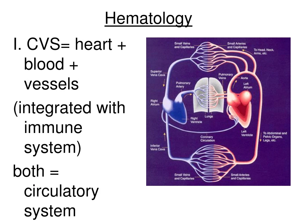
I. CVS= heart + blood + vessels (integrated with immune system) both = circulatory system. Hematology. II. Functions of blood;. transport gases, nutrients, hormones, metab. wastes reg. pH + ion conc. of interstitial fluid (electrolytes) restricts fluid loss at injury

Hematology
E N D
Presentation Transcript
I. CVS= heart + blood + vessels (integrated with immune system) both = circulatory system Hematology
II. Functions of blood; • transport gases, nutrients, hormones, metab. wastes • reg. pH + ion conc. of interstitial fluid (electrolytes) • restricts fluid loss at injury • defends against toxins and pathogens • stabilizes body temp.
III. Characteristics of blood • temp= 100 degrees F • pH= 7.35-7.45 (narrow range) • viscosity= 5 times more than water • amount= 7% of body weight in kg
IV. Composition; fluid CT • fluid is called plasma • cells (formed elements) are; • RBC- erythrocytes • WBC- leukocytes • Platelets – cell fragments • Hematocrit- shows % of solid elements • Centrifuged tube shows parts • (refer to plate)
V. Plasma characteristics; • straw colored • 46-63% of whole blood • 92% water • contains electrolytes and metabolites • contains 3 types of proteins • albumins- 60% of proteins; combines w/ and transports fatty acids and hormones, contributes to osmotic pressure • globulins- 35%; ex. Immunoglobulins(antibodies), transport globulins • fibrinogen- 4%; involved in clotting, blood w/o it is called serum
VI. Erythrocytes; • - made in red bone marrow • -1/3 of all cells in body • very specialized • shape is biconcave disc (p. 629) • 1. increases SA:V • 2. stackable to form rouleaux • 3. flexible due to spectrin • enucleated, no mitosis • no mitochondria or ribosomes • don’t use O2 (use anaerobic resp.) • packed with hemoglobin (Hb) • Life span 120 days
4 aa chains –2 alpha + 2 beta each chain wraps around a heme pigment heme will chem. comb. w/ oxygen Hb + O2 HbO2 Oxyhemoglobin makes bl. bright red O2 can disassociate making deoxyhemoglobin and bl. becomes burgundy VII. Hemoglobin (p. 630)
VIII. Recyclying RBCs –p. 632 1. RBCs become become engulfed by phagocytes of liver, spleen and bone marrow. - alpha and beta chains broken down into amino acids and metabolized or released into bl. -heme stripped of its iron and becomes biliverdin (green) 2 . biliverdin becomes bilirubin (yellow) 3. Bilirubin transported to liver to be excreted in bile. -.Jaundice develops when bile can’t be secreted. 2. Fe carried via transferrin to marrow to be recycled 3. . Excess Fe stored in ferritin in liver
IX. Hematopoiesis- production of cells - • refer to p. 643 • erythropoiesis- RBC formation
X. WBC’s • – most work outside the bl. stream inside tissues or in lymph organs • -2 groups- see plate A. Granular- (-phil) neutrophil- 50-70% eosinophil- 2-4% (stain red) basophil- <1% (stain blue) B. Agranular- (-cyte) lymphocyte- 20-30% monocyte- 2-8%
XI. WBC characteristics; • Ameboid movement • Diapedesis • Chemotaxis • Phagocytosis
Monocytes • largest WBC • spherical • tissue macrophage • largest component of pus
Neutrophils • lobed nucleus,polymorphs • pale colored granules • very mobile, fast • first to injury site • 10 hour life span • “eat” bacteria – form pus • die and release chemicals to attract more cells
Eosinophils • “eat” cells marked with antibodies • granules contain histamine • respond in allergic reactions • defend against parasitic infestations • (ex. hookworm and tapeworm)
Basophils • granules filled with heparin and histamine • respond in inflammation
Lymphocyte • large nucleus with little halo of cytoplasm • immune response • types of lymphocytes • (most not in bl.v.) • T cells- attack foreign cells • B cells- produce antibodies • NK cells- destroy abnormal cells
XII. Platelets- thrombocytes • (not complete cells) p. 643 • -produced by megakaryocyte in bone marrow • -responsible for hemostasis (clotting
XIII. Hemostasis- p. 647 1. Vascular phase – immediate contraction of bl.v 2. Platelet phase 0-30 secs a. damaged cells release chemicals b. damaged cells become sticky and contract c. platelets arrive at wound site and stick to damaged areas forming a “plug” 3. Coagulation phase 30 secs-8 min a. coagulation begins involving blood proteins called clotting factors (30 sec after (work in a chain reaction and need Ca+ and K+ to work) refer to p. 648 b. prothrombin forms thrombin enables fibrinogen to form fibrin c. prostaglandins released form tissues attract more platelets and clot forms (aspirin inhibits prostaglandin production) d. clot dries on surface and forms scab which pulls wounded tissue together
hemolytic anemia in the newborn • Occurs when a fetus is Rh+ and mother is Rh− • the mother will produce antibodies against the fetal antigen when blood is exchanged during birth. • complicates future pregnancies, because her antibodies will enter the fetal circulation system and react with fetal blood, causing hemolysis. • Treatment= Anti-Rh+ antibody is injected into the mother after her first delivery. -destroys the fetal Rh+ cells in the mother's circulation preventing Rh +antibody production in the mother -next child will not be at risk for hemolytic anemia.