Download
1 / 105
1.05k likes | 1.08k Views
Explore the skeletal structure, bone development, and microscopic features of long bones. Learn how bones grow, their classification, and the functions they serve in the body. Discover the intricate processes of bone formation and the importance of bone homeostasis.

E N D
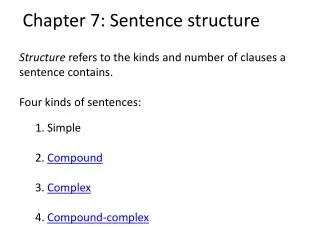
Skeletal Structure Chapter 7
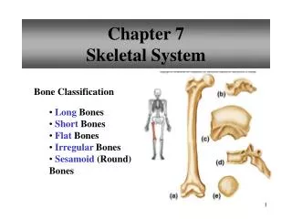
Bone Classification • Bones are grouped according to their shapes: • Long -femur • Short –tarsal bone of ankle • Flat –parietal bone of skull • Irregular –vertebra of backbone • round (sesamoid) -patella
Parts of a long bone • Epiphyses at each end are covered with articular cartilage and articulate with other bones. • Shaft is called diaphysis- it contains a medullary cavity filled c marrow • Bone is covered by periosteum • Compact bone has a continuous matrix with no gaps • Spongy bone has irregular interconnecting spaces between bony plates
Microscopic structure • Compact bone contains osteons OR osteocytes (these are called bone cells) cemented together. • Look p 198 • Central canals contains blood vessels that nourish the cells of ostoens. • Diffusion from the surface of the thin bony plates nourishes cells of spongy bones.
Compact Bone • Surrounds the spongy bone, with a continuous matrix, no gaps, tightly packed tissue
Bone Development and Growth • The skeletal system begins to form during the first few weeks of prenatal dev and cont to grow into adulthood • Bones form by replacing existing connective tissue in one of two ways: p. 201 chart • 1. intramembranous bones -some bones originate within sheetlike layers of connective tissues • 2. endochondral bones -others begin as masses of cartilage that are later replaced by bone tissue
Organic • Give bone a certain degree of flexibility
Inorganic • Give bone its hardness and durability
Intramembranous Bones • Certain flat bones of the skull are intramembranous bones. • They develop from layers of connective tissues. • Osteoblasts within the membranous layers from bone tissue. • Mature bone cells are called osteocytes. • Primitive connective tissue gives rise to the periosteum.
Endochondral Bones • Most of the bones of the skeleton are endochondral. • They develop as hyaline cartilage that is later replaced by bone tissue. • Primary ossification centers appear in the epiphyses. • An epiphyseal plate remains between the primary and secondary ossification centers.
Growth at the epiphyseal plate • An epiphyseal plate consists of layers of cells: resting cells, young dividing cells, older enlarging cells, dying cells. • The epiphyseal plates are responsible for lengthening. • Long bones continue to lengthen until the epiphyseal plates are ossified. • Growth in thinckness is due to intramembranous ossification beneath the periosteum. • The action of osteoclasts forms the medullary cavity.
Homeostasis of bone tissue • Ososteoclast and osteoblast continually remodel bone. • The total mass of bone remains nearly constant.
In other words: • Bones grow in length and ossify (harden) from the center of the diaphysis (the shaft) toward the epiphyseal (ends) extremities epiphyseal plate (metaphysis) • It lengths in the middle so as not to disturb the joints that are already formed • Look at xray p. 203 read purple box
What is an Osteoblast? • Bone cell that deposit new bone material in central part of bone dissolves to form the medullary canal. • This gets bigger as the diameter of bone increases • A bone forming cell
What are Osteoclasts? • Large bone cells that secrete enzymes that dissolve or digest the bone material (to form medullary canal) other minerals are broken down by these cells and absorbed by surrounding fluid • A cell that erodes bone
When does growth stop? • The length of the bone shaft cont to grow until all epiphyseal (ends) cartilage is ossified • Average growth of female cont about 18 yr • Average gr of males 20-21 yr • New growth can occur in a broken bone @ anytime
Factors affecting bone development, growth, and repair • Deficiencies of vitamin A, C, or D result in adnormal development. • Insufficient secretion of pituitary growth hormone may result in dwarfism; excessive secretion may result in gigantism, or acromegaly. • Deficiency of thyroid hormone delays bone growth. • Male and female sex hormones promote bone formation and stimulate ossification of the epiphyseal disks.
Support and Protection • Bones shape form body structure • Bones support and protect softer, underlying tissues.
Body Movement • Bones and muscles function together as levers. • A lever consists of a rod, a pivot, a resistance, and a force that supplies energy.
Blood Cell Formation • At the different ages , hemopoiesis occurs in the liver, the spleen, and the red bone marrow. • Red marrow houses developing red blood cells, white blood cells, and blood platelets.
Inorganic Salt Storage • The intercellular material of bone tissue contains large quantities of calcium phosphate in the form of hydroxyapatite. • Bone stores small amounts of sodium, magnesium, potassium, and carbonate ions. • Bone tissues may accumulate lead, radium, or strontium.
Number of Bones • Usually a human skeleton has 206 bones, but the number may vary. • Extra bones in sutures are called sutural bones.
Divisions of Bones • The skeleton can be divided into axial and appendicular portions. • The axial skeleton consists of the skull, hyoid bone, vertebral column, and thoracic cage.
Axial skeleton--support & protect organs of head, neck & truck • 1. skull-cranium and facial bones • 2. hyoid bone- in neck b/w lower jaw & larynx *only bone of the body that does not articulate with any other bones— • How does the hyoid bone remain attached?—it is fixed by muscles & ligaments & supports tongue & is an attachment for certain muscles that move tongue during swallowing • 3. Vertebral column-sacrum (part of pelvis) & coccyx • 4. Thoracic Cavity- 12 pairs ribs & sternum • 5. Middle ear bones- malleus (hammer), incus (anvil), stapes (stirrup)
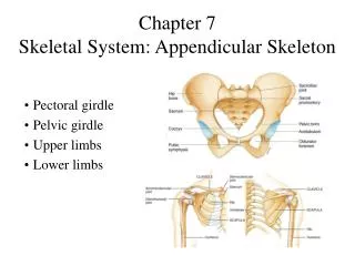
Appendicular Skeleton-consists of bones of upper/lower ext & the bones that anchor the limbs to axial skeleton • 1. Pectoral girdle- formed by scapula & clavicle Connects upper ext to axial skeleton • 2. Upper limbs- humerus, radius, ulna, carpal, metacarpal, phalanx or phalanges • 3. Pelvic girdle- 2 os coxae-hip bones attached to each other anteriorly & to sacrum posteriorly- • Ilium-top • Ischium-forms loop • Pubis-front
…cont app skeleton • 4. Lower limbs- • Legs-femur, tibia, fibula, patella • Feet- ankle-tarsals, metarsals, phalanges
Cranium • The cranium encloses and protects the brain and provides attachments for muscles. • Some cranial bones contain air filled sinuses that help reduce the weight of the skull.
8 bones of the Cranium • 1. Frontal (1)- forms forehead • 2. Two Parietal- roof of cranium • 3. Two temporal- side of skull • 4. Occipital (1)- back of skull • 5. Ethmoid (1)- forms part of roof & wall of nasal cavity-sinus • 6. Sphenoid (1)- forms part if base of cranium, sides of skull, sides of orbits-sinus
Facial Skeleton • Facial bones form the basic shapes of the face and provide attachments for muscles.
14 Facial Bones p. 224 T7.7 • 1. Mandible-lower jaw • 2. Maxilla-two bones forming upper jaw • 3. Zygomatic- two check bones • 4. Nasal- five bones in upper part of nose- vomer (1), inferior nasal concha (2), nasal (2) • 5. Lacrimal- two bones at inner aspect of eyes • 6. Palatine- two bones of hard palate or roof of mouth, behind maxilla
Infantile Skull • Incompletely developed bones, connected by fontanels, enable the infantile skull to change shape slightly during childbirth. • Proportions of the infantile skull are different from those of an adult skull, and its bones are less easily fractured.
Page 225 Vertebral Column
What do the Vertebra do? • Extends from skull to pelvis • Composed of many (26) vertebrae that are separated by masses of fibrocartilage called intervertebral disks (shock absorbers) and are connected to one another by ligaments. • Supports the head & trunk of the body • Flexible, causes movement-bending, turning, rotating on central axis. • Protects spinal cord
Turn to p. 226 • 7 cervical • 12 thoracic • 5 lumbar • ****remember these like the times when you eat!!!
7 Cervical Vertebrae-NECK • Cervical vertebrae comprise the bones of the neck. • Transverse processes have transverse foramina. • The atlas (the first vertebra) supports the head. • The dens of the axis (the second vertebra) provides a pivot for the atlas when the head is turned from side to side.
12 Thoracic Vertebrae-RIBS • Thoracic are larger than cervical vertebrae. • Their long spinous processes slope downward, and facets on the sides of bodies articulate with the ribs.
5 Lumbar vertebrae-WAIST • Vertebral bodies of lumbar vertebrae are longer and stronger. • Their transverse processes project posteriorly at sharp angles, and their spinous processes are directed horizontally.