Download

1 / 29
290 likes | 308 Views
This introduction to fluoroscopic investigations of the gastrointestinal tract covers the anatomy, function, and examination methods. It also discusses radiographic contrast and contrast media used in GIT exams.

E N D
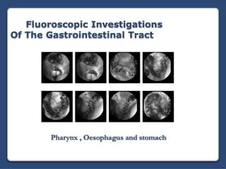
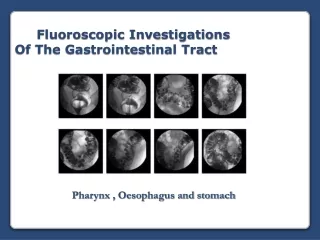
Fluoroscopic Investigations Of The Gastrointestinal Tract ( Introduction)
Learning objectives • With the end of this lecture the student will be able to: • Describe the anatomy of the digestive system, to include the pharynx, esophagus ,stomach, , large intestine ,and accessory organs • Discuss the orientation of the stomach according to body habitus, physical position and respiration • Identify the function of each of the structures of the digestive system • Describe room preparation and identify supplies needed for the examinations of the digestive system • Identify methods of evaluating the digestive system • State the criteria to evaluate the digestive system radiographs in term of positioning ,image quality, radiographic anatomy ,and pathology • Explain different types of contrast media used in GIT exams • Explain pharmacological agents used in GIT exams • Differentiate between Retrograde and Antegrade GIT studies
References • Radiographic procedures: By Stephen Chapman • Positioning in Radiography: By k.C.Clarke. • Text book of radiographic positioning and related anatomy; by • Kenneth L.Bontrager 6th edition. • Websites • http://www.e-radiography.net/
Basic Anatomy • The Digestive system Consists of: • Mouth • Pharynx • Oesophagus • Stomach • Small Intestine • Large Intestine • Rectum & Anus • Accessory organs include • Tongue and Teeth • Salivary glands • Pancreas • Liver • Gall bladder & biliary tract
Physiology of the digestive system Ingestion Digestion Absorption Elimination
Investigations of the digestive system • Plain film Investigation • Barium Investigations • Radionuclide Imaging • Computerized Tomography • Magnetic Resonance Imaging • Angiography • Ultrasound • Non- Radiographic Investigations (Endoscopy)
Barium Investigations • Wide range of investigations can be performed • Can incorporate plain film & fluoroscopic equipment • Involves the use of contrast media • Can involve the use of pharmacological agents • What is the role of the Radiographer?
Short (high) Long (low) Radiographic Contrast vs. Contrast Media • Radiographic Contrast: Difference between adjacent densities in a radiograph. • The films or images have different levels of density – different shades of gray • MAJORDIFFERENCES • SLIGHTDIFFERENCES
Radiographic Contrast vs. Contrast Media • CONTRAST • X-RAY DYE • DYE • Contrast Media: Diagnostic agents that are instilled into body orifices or injected into the vascular system, joints, and ducts to enhance subject contrast in anatomic areas where there is low subject contrast.
Purpose of Contrast Media • To enhance subject contrast or render high subject contrast in a tissue that normally has low subject contrast.
Contrast Media Properties • able to show organ better • physiologically • no permanent alteration of organ • non toxic and able to be eliminated / excreted Negative contrast • Radiolucent e.g. AIR • Low atomic # material • Black on film Positive contrast • Radiopaque e.g. BARIUM • High atomic Number • White on film
Barium Sulfate: BaSO4 Contrast Media for GIT Exams • High atomic number • Not soluble in water = suspension • Used to coat the lining of organs • Supplied in different thicknesses • Used • Esophogram, UGI, Small Bowel, Large Bowel Contraindications • perforations of GI tract • proximal to an obstructed bowel Precautions • adequate hydration post examination
Contrast Media for GIT Exams Why use Barium Sulphate? • It has a high atomic number (Z=56) • Non-toxic • Relatively cheap • body cannot metabolize BaSO4
Contrast Media for GIT Exams Gastrografin or Hypaque • High atomic # • Close to iodine • Water soluble • Similar usage as Barium • Water soluble, safe in the abdominal cavity • Safe to use if perforation is suspected
Pharmacological agents • Buscopan • Glucagon • Maxalon • Carbex • Methyl cellulose • Why are they given?
GI Contrast Studies • Antegrade studies (with the normal flow) • esophagus, stomach, small bowel • Contrast (Barium / barium + air /Oral iodine solution BaSO4 Only BaSO4 + Air
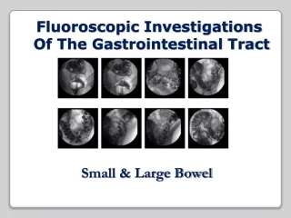
GI Contrast Studies • Retrograde studies (against the flow) • Colon ( Large Bowel) • Contrast barium / barium + air Barium + Air Barium Only
GI Contrast Studies (Preparation) Common patient preparation aspects for barium investigations What would you check? • Identification, request form. • Pregnancy check (10 day rule) • Review of previous examinations • Allergy check • Abdominal preparation • ?Diabetic patients (adjustment in appointment time & preparation prior to examination) • Psychological preparation for examination
GI Contrast Studies (Preparation) Summary of abdominal preparation ( Adults)
GI Contrast Studies (Preparation) Summary of abdominal preparation ( Paediatrics)
GI Contrast Studies (Breathing Instructions) • To reduce the stress of the abdomen that could lead to involuntary motion, • all radiographs are obtained during suspended respiration. • Special breathing techniques are used to increase the Intrathoracic and • intra abdominal pressure
GI Contrast Studies Equipment consideration • Tilting table (+/-90 degrees) • Rapid film recording facilities • Special feeding accessories • Fluoroscopic unit (normally digital) • Additional over couch tube • Lead aprons and gloves • Sponges and other supports
GI Contrast Studies (Radiation protection) • ALARA principle (Minimise dose to patient /staff) • Utilise pulsed fluoroscopy where appropriate • Record patient dose & imaging times • Record any faults with equipment / regular QA • Practical collimation • Adhere to clinical protocols (minimum no of films / dose) • Always read patient history / notes
GI Contrast Studies (Plain Films) Before barium Studies Plain Films are required To Assess:- • Trauma • Small / Large Bowel Obstruction • Constipation • Palpable Mass • Acute Gastro-intestinal Bleeding • Perforation (include. Chest radiograph)
GI Contrast Studies(Plain Films) Plain films showing Obstruction