Download
1 / 36
600 likes | 2.1k Views
Fluoroscopic Investigations Of The Gastrointestinal Tract. Pharynx , Oesophagus and stomach. References. Radiographic procedures: By Stephen Chapman Positioning in Radiography: By k.C.Clarke. Text book of radiographic positioning and related

E N D
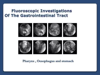
Fluoroscopic Investigations Of The Gastrointestinal Tract Pharynx , Oesophagus and stomach
References • Radiographic procedures: By Stephen Chapman • Positioning in Radiography: By k.C.Clarke. • Text book of radiographic positioning and related • anatomy;bykenneth L.Bontrager. • Websites • http://www.e-radiography.net/
Learning Objectives • With the end of these lectures the student will be able to: • List common indications for ordering Ba swallow /meal exam • Explain Ba swallow/meal exam limitations • Explain the contraindications for using barium sulphate in the examination of the oesophagus and stomach. • Describe the anatomy of the oesophagus and stomach and explain their function • Describe room preparation and identify supplies for Ba swallow and barium meal series • Describe how to perform barium swallow / meal • Explain patient care, after completing the barium procedures • Critique Ba swallow /meal radiographs in term of positioning ,image quality, radiographic anatomy ,and pathology
What is the function of esophagus? Transport of food by peristalsis.
Barium Swallows –Indications • Pain on swallowing • Fistulae between trachea & oesophagus • (non-ionic c/a preferred) • Assessment of action of oesophagus following a stroke • Oesophageal varices / Diverticula • As part of a barium meal investigation • Dysphagia • Carcinoma / obstruction /Hiatus hernia • Hemetemesis
Barium Swallow - Technique • a series of plain films or uses fluoroscopy to identify any pathology • Patient is placed in the erect RAO position • Ample mouthful of barium is swallowed & spot films are taken (rapid sequence) • Spot films of the upper & lower oesphagus are taken • May need rapid serial radiography sequence
Barium Swallow - Technique • Following Barium Swallow upper GI series may performed to diagnose pathology in the, stomach, and duodenum Limitations • Not good for evaluating small ulcers • Not specific for diagnosis of esophagitis
Barium Swallow (Normal Films)
Figure2 Figure1 Figure1: Shows the lower end of a normal esophagus with a smooth connection between the lower esophagus and stomach. Figure 2: Shows the lower end of the esophagus with a small hiatus hernia, which occurs when a small portion of the stomach pushes up into the chest.
Barium Swallow AP RAO
Aftercare of the patient • Patient given tissue to wipe & clean mouth • Patient aware of where & when to obtain results. • Patient given the chance to ask any questions. • The patient should drink plenty of fluids and • may need a laxative after the test because • the barium can be constipating
Barium Swallow (Pathology Films)
ACHALASIA Distended esophagus with distil stricture due to Achalasia - Failure of distil sphincter to relax – causing obstruction.
Esophageal Spasm Strictures
Tracheo - oesophageal fistula Normal Swallow Leaks of contrast into the trachea
Stomach Barium meal
Stomach Anatomy • J-Shaped • Continuous with Oesophagus & duodenum • Three sections • Fundus • Body • Pyloric Antrum
Barium Meal Indications • Dyspepsia / reflux / Upper abdomen pain/ Nausea/ Weight loss • Fullness or distension • Peptic ulceration (defects in mucosa extending through muscularis mucosae) • Gastritis ( Inflammation of the stomach) • Polyps • Upper abdominal mass • Gastrointestinal haemorrhage • Pyloric / cardiac stenosis • Hiatus hernia ( Slipping of the upper portion of the stomach through the oesophageal hiatus • Partial bowel obstruction • Assessment of site of perforation (What type of contrast to use?) • Contra-indications: • Complete bowel obstruction
Barium meal Contrast media & patient preparation • High density, low viscosity barium • Nil orally for 6 hours prior • Explanation of procedure • Physical & psychological preparation • No smoking (>gastric motility) • Check for contra-indications to pharmacological agents • ( What are the contra indication for Buscopan?)
Barium Meal Investigation • Can perform double (CO2 & Barium) or single contrast examinations • Single contrast examinations are used in paediatrics & grossly ill patients • Double contrast examinations - demonstrate mucosal pattern • Equipment should contain ability to perform spot film images.
Barium meal - Technique • Gas producing agent swallowed (eg. Carbex) • Patient drinks barium whilst lying on left side • Patient lies supine & slightly on their right side • Check for reflux • Smooth muscle relaxant given to the patient • Buscopan (20mg iv) or Glucagon (0.3mg iv) • Patient rolls onto their right side & quickly over in a complete circle - finish in a RAO position • This has the effect of coating the gastric mucosa with barium
Barium meal - Typical film series RAO Stomach and C-loop of the duodenum with duodenal bulb in profile
Barium meal - Typical film series PA (Prone) Duodenal loop + duodenal with body and pylorus filled with barium
Barium meal - Typical film series Right lateral Retro gastric space
Barium meal - Typical film series AP (supine) Entire stomach and duodenum + Fundus of stomach filled with barium
Barium meal - Typical film series LPO Duodenum Bulb without superimposition with the pylorus + Fundus of stomach filled with barium LAO Lesser curve Prone , RAO, LAO , Supine, Erect Duodenal Cap series Note : In the erect position the Fundus of the stomach is filled with air
Barium meal ( Normal anatomy) • (3) greater curvature (4)lesser curvature(5)fundus (6)small bubble of gas. (7)pyloric region(8)second part of the duodenum
( Pathology) PYLORIC STENOSIS
( Pathology) GASTRIC CARCINOMA
( Pathology) *Note distended distil esophagus with herniation of gastric fundus into chest through esophageal hiatus. DIAPHRAGM Normal Hiatus Hernia
( Pathology) DUODENAL ULCER
Any Questions? Thank you