Download

1 / 109
1.11k likes | 1.31k Views
Neoplasms of the Gastrointestinal Tract. Dr. M Jeffers Consultant Histopathologist AMNCH, Tallaght. Objectives: GI tumour pathology. tumour nomenclature and classification tumour aetiology and pathogenesis precursor lesions mechanisms of carcinogenesis in different tissues

E N D
Neoplasms of the Gastrointestinal Tract Dr. M Jeffers Consultant Histopathologist AMNCH, Tallaght
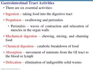
Objectives: GI tumour pathology • tumour nomenclature and classification • tumour aetiology and pathogenesis • precursor lesions • mechanisms of carcinogenesis in different tissues • potential points for prevention / modification • tumour growth and behaviour • invasion • metastasis • tumour staging • staging systems • prognostic factors AMNCH Tallaght
Useful links • http://medstat.med.utah.edu/WebPath/webpath.html • www.bmj.com : collected resources • ABC of colorectal cancer • ABC of the upper gastrointestinal tract: cancer of the stomach and pancreas AMNCH Tallaght
Neoplasms • Definition • Epidemiology, Aetiology, Pathogenesis • Morphology: Gross / Microscopic • Clinical presentation and outcome • Prognosis AMNCH Tallaght
Nomenclature and Classification • Nomenclature / Classification by histogenesis: • Epithelial • Mesenchymal • Neuroendocrine • Haematolymphoid • Melanocytic AMNCH Tallaght
Nomenclature and Classification • Epithelial tumours: • Benign: “adenoma” • Malignant: “carcinoma” • Adenocarcinoma Squamous carcinoma Small cell carcinoma AMNCH Tallaght
Nomenclature and Classification • Neuroendocrine tumours: • Benign: “carcinoid” • Malignant: neuroendocrine carcinoma • small cell carcinoma AMNCH Tallaght
Nomenclature and Classification • Mesenchymal tumours: • Benign: (tissue)oma lipoma, leiomyoma etc • Malignant: (tissue)sarcoma liposarcoma, leiomyoma etc • Gastrointestinal stromal tumour (GIST): benign • low malignant potential • malignant AMNCH Tallaght
Nomenclature and Classification • Haematolymphoid tumours: • Bone marrow neoplasm (+/- circulating cells): leukaemia • Lymph node / extranodal lymphoid cell tumour: lymphoma AMNCH Tallaght
Neoplasms of the Gastrointestinal Tract • Concepts: • Carcinogenesis: • Metaplasia/Dysplasia/Carcinoma sequence • Adenoma/Carcinoma sequence • Differentiation • Invasion and Metastasis • Tumour staging AMNCH Tallaght
Tumour Aetiology and Pathogenesis: Carcinogenesis • Various mechanisms operative in carcinogenesis in the GI tract: • Intraepithelial neoplasia – Dysplasia – Carcinoma • Metaplasia – Dysplasia - Carcinoma • Adenoma - Carcinoma sequence • Replicator error phenotype AMNCH Tallaght
Intraepithelial neoplasia – carcinoma sequence • Multistage process of carcinogenesis from • normal through • low grade intra-epithelial neoplasia (dysplasia) (RR 2.2) through • high grade intra-epithelial neoplasia (dysplasia) (RR >60) to • invasive carcinoma. • Progressive development of architectural and cytological abnormality • Progressive increase in relative risk of invasive carcinoma • Useful model is squamous cell carcinoma of oesophagus. AMNCH Tallaght
Intraepithelial neoplasia – carcinoma sequence: Oesophageal cancer • Morphologic abnormality in dysplasia –carcinoma sequence in oesophageal mucosa reflects underlying genetic abnormality. • p53 mutation • low grade • LOH 3p14 (FHIT) fragile histidine triad, tumour suppressor gene • Cyclin D1 overexpression (11q13), 3p21 LOH • C-myc, EGFR, HST1 overexpression invasive carcinoma AMNCH Tallaght
Intraepithelial neoplasia – carcinoma sequence Normal oesophageal squamous mucosa High grade squamous dysplasia (carcinoma in-situ) Invasive squamous cell carcinoma p53 mutation FHIT LOH CYD1 c-myc, EGFR, HST1 AMNCH Tallaght
Metaplasia – Dysplasia – Carcinoma Sequence • Multistage process of carcinogenesis from • Normal through • Metaplastic columnar epithelium through • Dysplasia in metaplastic epithelium • low grade • high grade to • Invasive carcinoma • Useful model is adenocarcinoma of oesophagus AMNCH Tallaght
Metaplasia – Dysplasia – Carcinoma sequence: Oesophageal cancer • Barrett’s oesophagus = columnar lined oesophagus • Replacement of the squamous lining of the distal oesophagus by glandular mucosa in response to injury, most frequently gastro-oesophageal reflux • metaplasia to gastric and intestinal type epithelium • Stepwise progression of dysplasia in metaplastic epithelium leads to invasive malignancy: architectural and cytological abnormalities develop. • Morphologic changes reflect underlying molecular genetic / cell cyle regulatory abnormalities AMNCH Tallaght
Metaplasia – Dysplasia – Carcinoma sequence: Oesophageal cancer • Normal • Metaplasia • FHIT alterations, CDKN2A hypermethylation • Dysplasia • rab11 abnormalities • Low grade • Ki67 abnormality • High grade APC mutation • p53 mutation (different to scc) • Invasive carcinoma c-erbB2, EGFR AMNCH Tallaght
Metaplasia – Dysplasia – Carcinoma sequence: Oesophageal cancer Normal oesophageal squamous mucosa Barrett’s oesophagus: metaplastic columnar and intestinal mucosa AMNCH Tallaght
Metaplasia – Dysplasia – Carcinoma sequence: Oesophageal cancer Barrett’s oesophagus: low grade dysplasia Invasive adenocarcinoma Barrett’s oesophagus: high grade dysplasia FHIT Ki67 p53 APC AMNCH Tallaght
The metaplasia dysplasia sequence: significance • Intestinal metaplasia in the oesophagus is a marker of increased adenocarcinoma risk • Risk of carcinoma is greatest with high grade dysplasia: concurrent carcinoma in up to 50% • Many cases of high grade dysplasia will progress to invasive carcinoma over time • Diagnosis of high grade dysplasia: rebiopsy to exclude invasion • consider surgery • Diagnosis of metaplasia: treat underlying conditions (GORD) • surveillance endoscopy to identify dysplasia • aiming to prevent cancers / detect cancer early AMNCH Tallaght
Metaplasia – Dysplasia – Carcinoma sequence: Gastric cancer • Chronic atrophic gastritis is a major risk factor for gastric carcinoma (intestinal type) • Risk of malignant transformation is strongly linked to intestinal metaplasia and intra-epithelial neoplasia (dysplasia) • Variety of processes may lead to atrophic gastritis: • Helicobacter gastritis • Auto-immune gastritis • Metaplasia – dysplasia sequence acts as a final common pathway in development of malignancy AMNCH Tallaght
H Pylori infection Nitrate reductase Gastritis Diet iNOS expression Nitrite Acid NO N2O3 Ascorbic acid Cell damage (DNA, lipids, mitochondria etc) Nitrosamines Repair Mutation Apoptosis Cancer Metaplasia / dysplasia Atrophic gastritis AMNCH Tallaght
Metaplasia – Dysplasia – Carcinoma sequence: Gastric cancer • Chronic gastritis • Intestinal metaplasia • Dysplasia • low grade • high grade • Invasive carcinoma AMNCH Tallaght
Metaplasia – dysplasia - carcinoma sequence in the stomach Stomach: chronic gastritis Stomach: normal Stomach: atrophy and intestinal metaplasia AMNCH Tallaght
Metaplasia – dysplasia - carcinoma sequence in the stomach Stomach: high grade dysplasia Stomach: low grade dysplasia Stomach: invasive adenocarcinoma AMNCH Tallaght
The metaplasia dysplasia sequence: significance • Most intestinal type gastric cancers develop on a background of atrophy, metaplasia and dysplasia. • Predisposing factors for atrophy and metaplasia are known: • chronic HP gastritis, auto-immune gastritis etc. • Treatment of predisposing conditions, endoscopic surveillance of patients with documented metaplasia: strategies to reduce risk, detect cancer early. AMNCH Tallaght
Colorectal carcinogenesis: the adenoma – carcinoma sequence • Various epidemiological associations in colorectal carcinoma point to the important role of the adenoma as the precursor lesion of many cancers • Morphological progression in colorectal carcinoma: • Aberrant crypt focus dysplastic ACF with APC mutation inactivation • Adenoma proto-oncogene activation: c-myc, ras • low grade dysplasia suppressor gene inactivation: p53, p21, bax • high grade dysplasia • other abnormalities: BRCA, DCC, E-cad • Carcinoma AMNCH Tallaght
Carcinoma Methylation Defect p53 K-Ras Smad4 ?? Normal Epithelium Early Adenoma Intermediate Adenoma Late Adenoma Metastasis APC Carcinoma MSI + Mismatch repair defect TGF, BAX ?? Genetic Model for Sporadic Colorectal Carcinogenesis AMNCH Tallaght
Adenomatous polyps of the colon • Neoplastic polyps, precursors of carcinoma • Architectural patterns: • Tubular/Villous/Tubulovillous • Gross presentation: • sessile/pedunculated/flat adenoma • Risk of malignancy • size (ras), architecture, dysplasia AMNCH Tallaght
Colon: tubular adenoma AMNCH Tallaght
normal epithelium adenomatous epithelium AMNCH Tallaght
Colon: villous adenoma AMNCH Tallaght
Colon: villous adenoma AMNCH Tallaght
Polyposis syndromes • Sydromes characterised by multiple polyps in colorectum +/- elsewhere in GIT • FAP: 5q21 defect, multiple polyps, 100% lifetime risk of carcinoma • HNPCC: MSH gene defect, fewer polyps, typical pattern of tumours • Gardner: variant of FAP with skin and other lesions • Turcot: association with CNS neoplasms AMNCH Tallaght
Table 1. Intestinal polyposis syndromes Location Location of Location ofSyndrome Inheritance Type(s) of polyp of polyp Gl cancer extraintestinal tumorsFamilial polyposis AD Adenoma, FGP C, SI, ST C, SI Nonecoli (familial adenomatous polyposis) Gardner’s AD Adenoma; FGP, C, SI, ST C, SI Lipoma; fibroma; desmoid lymphoid tumor; dental cysts;osteoma; carcinoma ofthyroid and adrenal glandsTurcot’s* AD Adenoma C C Central nervous system tumorsPeutz-Jeghers AD Hamartoma C,SI,ST C,SI,ST Cancers of the breast,ovary, uterus, and testis Juvenile polyposis AD Juvenile C,SI,ST C,SI,ST None(one-third of cases) Cowden’s AD Hamartoma; C,SI None Facial trichilemomas;inflammatory; oral mucosal papillomas; ganglioneuroma; carcinoma of thyroidlipoma; lymphoid gland and breastCronkite-Canada None Juvenile-like C,SI,ST Colon (rare) NoneAttenuated AD Adenoma; FGP C,SI,ST C,SI,ST Nonefamilial polyposis coli** Hereditary flat AD Adenoma; FGP C,SI,ST C,SI,ST NoneAdenoma syndrome** Muir Torre syndrome AD Adenoma C C Skin cancer: basal cell;squamous cell; sebaceouscarcinomaHereditary mixed AD Adenoma; juvenile; C C Nonepolyposis syndrome hyperplastic GI, gastrointestinal, AD, autosomal dominant, FGP, fundic gland polyp; C, colon; SI, small intestine; ST, stomach. *Some cases of Turcot’s syndrome have mutations in APC gene, Some cases of Turcot’s syndrome have germline mutations in mismatch repair genes (hMLH1 and hPMS2). **Attenuated familial polyposis coli and hereditary flat adenoma syndrome most likely are the same disorder, but they are listed separately in this table. Attenuated familial polyposis coli and hereditary flat adenoma syndrome are considered variants of familial polyposis coli. AMNCH Tallaght
Familial adenomatous polyposis (FAP) • Autosomal dominant condition characterised by numerous adenomatous polyps and high risk of progression to adenocarcinoma • Germline mutation in APC gene (5q21-22) which encodes a 2843 aa polypeptide which acts as a negative regulator in the Wnt signalling pathway, regulates cellular -catenin concentration • Lack of functional APC leads to accumulation of -catenin and consititutive expression of c-myc and cyclin D1 AMNCH Tallaght
Colon: familial adenomatous polyposis AMNCH Tallaght
Adenoma carcinoma sequence: significance and implications • Mechanism underlying most colorectal carcinomas • Identifiable precursor lesions in many cases: early detection of precursor lesions has obvious implications for early treatment and prevention of invasive disease • Identification of families with germ-line mutations and significant polyposis syndromes offers significant preventive opportunities • Tumour pathogenesis has direct implications on cancer management and prevention AMNCH Tallaght
Colorectal carcinogenesis: the mismatch repair defect pathway • Mechanism of carcinogenesis distinct from the adenoma – carcinoma sequence • Tumours characterised by extensive nucleotide insertions / deletions in repeated sequences in tumour DNA: microsatellite instability (MSI) / DNA replication error (RER) • Tumours with MSI are classified as MSI high-frequency (MSI-H) or • MSI low frequency (MSI-L) • These tumours arise through defects in the DNA mismatch repair mechanism, defects which may be sporadic or inherited • Some sporadic tumours are MSI-H but most MSI-H tumours characteristic of hereditary non-polyposis colorectal carcinoma (HNPCC) AMNCH Tallaght
HNPCC • HNPCC: a syndrome characterised by inherited defects in DNA repair due to germline mutations of the relevant genes: (hMLH1, hMSH2) • High frequency of colorectal carcinoma • Extracolonic tumours in endometrium, stomach, ovary, brain, skin, small bowel • Criteria for classification as HNPCC: Amsterdam criteria (+ revisions) • Classical tumour characteristics: right sided, large, mucinous, low stage AMNCH Tallaght
Hereditary Non-polyposis Colorectal Cancer • Criteria: • At least 3 relatives with a HNPCC associated cancer (colorectum/endometrium/small bowel/ureter/renal pelvis) • Ones should be first degree relative of other two • At least 2 successive generations involved • At least one tumour diagnosed before 50 yrs of age • Familial adenomatous polyposis should be excluded • Tumours should be verified by histopathological examination AMNCH Tallaght
MSI in colorectal cancer: diagnosis • hMSH2: normal hMSH2: tumour Normal tissue: hMSH2 expression Loss of expression in MSI-H tumour AMNCH Tallaght
Replicator error phenotype: implications • Identification of MSI-H tumours confers a high probability of an inherited cancer • Cancers can be screened for MSI status, confirmation of germ-line status required for definition of syndrome • Careful family history combined with tumour characteristics can identify at-risk groups • Early intervention and prevention opportunities • Tumour pathogenesis impacts directly on patient management and prognosis. AMNCH Tallaght
Carcinogenesis: review of mechanisms • Various mechanisms operative in carcinogenesis in the GI tract: • Intraepithelial neoplasia – Dysplasia – Carcinoma • Metaplasia – Dysplasia - Carcinoma • Adenoma - Carcinoma sequence • Replicator error phenotype Various mechanisms implicated at different sites Significance in terms of identification of precursor lesions, treatment and prevention of cancer AMNCH Tallaght
Differentiation • Differentiation refers to the degree of maturation of tumour cells / tissues: • what cell type / tissue type does the tumour grow in ? • how closely does the tumour reproduce normal tissue architecture ? • Tumours are described as: well differentiated • moderately differentiated • poorly differentiated • Differentiation is closely related to tumour grade: • well differentiated – low grade • poorly differentiated – high grade AMNCH Tallaght
Differentiation: colorectal carcinoma well differentiated moderately differentiated poorly differentiated AMNCH Tallaght
Neoplasms of the Gastrointestinal Tract • Classification of tumours: • Anatomical Location • Origin (Primary/Secondary) • Behaviour (benign/uncertain/malignant) • Histogenesis (epithelial/stromal/lymphoid etc) AMNCH Tallaght
Tumours of the oesophagus • Benign tumours rare: • Leiomyoma • Most are malignant and most are carcinomas • Squamous cell carcinoma • Adenocarcinoma AMNCH Tallaght