Download
1 / 56
600 likes | 811 Views
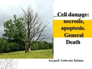
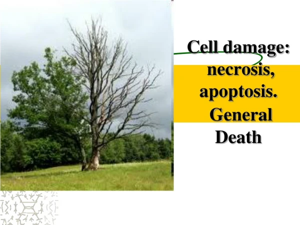
Necrosis and apoptosis. Objectives. Define necrosis and apoptosis List the different types of necrosis, examples of each and its features List the different conditions associated with apoptosis, its morphology and its mechanism Know the difference between apoptosis and necrosis. Objectives.

E N D
Objectives • Define necrosis and apoptosis • List the different types of necrosis, examples of each and its features • List the different conditions associated with apoptosis, its morphology and its mechanism • Know the difference between apoptosis and necrosis
Objectives • Define necrosis and apoptosis • List the different types of necrosis, examples of each and its features • List the different conditions associated with apoptosis, its morphology and its mechanism • Know the difference between apoptosis and necrosis
Cell Death • Death of cells occurs in two ways: • Necrosis--(irreversible injury) changes produced by enzymatic digestion of dead cellular elements • Apoptosis--vital process that helps eliminate unwanted cells--an internally programmed series of events effected by dedicated gene products
Objectives • Define necrosis and apoptosis • List the different types of necrosis, examples of each and its features • List the different conditions associated with apoptosis, its morphology and its mechanism • Know the difference between apoptosis and necrosis
Morphology of necrosis : • Cellular swelling or rupture • Denaturation and coagulation of cytoplasmic proteins • Breakdown of cell organelles • Breakdown of nuclear DNA
Patterns of Necrosis In Tissues or Organs • As a result of cell death the tissues or organs display one of these six macroscopic changes: • Coagulative necrosis • Liquifactive necrosis • Caseous necrosis • Fat necrosis • Gangrenous necrosis • Fibrinoid necrosis
Patterns of Necrosis In Tissues or Organs • Coagulative necrosis: • the outline of the dead cells are maintained and the tissue is somewhat firm. • Example: myocardial infarction • Liquefactive necrosis: • the dead cells undergo disintegration and affected tissue is liquefied. • Example: cerebral infarction.
Patterns of Necrosis In Tissues or Organs • 3. Caseous necrosis: • a form of coagulative necrosis (cheese-like). Example: tuberculosis lesions. • 4. Fat necrosis: • enzymatic digestion of fat. • Example: necrosis of fat by pancreatic enzymes. • 5. Gangrenous necrosis: • Necrosis (secondary to ischemia) usually with superimposed infection. • Example: necrosis of distal limbs, usually foot and toes in diabetes. • 6. Fibrinoid necrosis: typically seen in vasculitis and glomerular autoimmune diseases
Coagulative Necrosis Changes in the cytoplasm and the nucleus • Karyorrhexis • Karyolysis • Pyknosis
Necrosis of the liver induced by herpesvirus. Herpesvirus invades liver cell nuclei producing nuclear ('ground glass') mildly basophilic inclusions.
Acute renal tubular necrosis (ischemia) : increased eosinophilia and pyknosis in necrotic cells Normal Necrotic
Ischemic neuronal injury:acidophilic cytoplasm & nuclear pyknosis
… a mess - so many cells have died - the tissue is not recognizable. Nuclei have become pyknotic (shrunken and dark), undergone karorrhexis (fragmentation) and karyolysis(dissolution) Necrotic myocardium
Coagulative necrosis Kidney: ischemia and infarction (loss of blood supply and resultant tissue anoxia). Removal of the dead tissue leaves behind a scar
Coagulative necrosis: is due to loss of blood supply Infarcts (vascular distribution) are wedge-shaped with a base on the organ capsule. Spleen
Depending on circumstances: • necrotic tissue may be walled off by scar tissue, • totally converted to scar tissue, • get destroyed (producing a cavity or cyst), • get infected (producing an abscess or "wet gangrene"), • or calcify. • If the supporting tissue framework does not die, and the dead cells are of a type capable of regeneration, you may have complete healing. Remember: True coagulation necrosis involves groups of cells, and is almost always accompanied, by acute inflammation (infiltrate)
Liquefactive Necrosis Rate of dissolution of the necrotic cells is faster than the rate of repair.Usually results in an abscess secondary to bacterial infection. Liquefactive necrosis : hydrolysis of dead tissues or cells (rapidly destroyed by lysosomal enzymes from neutrophilic leukocytes (i.e., bacterial infections), or clostridia or snake poison. Liquefactive necrosis that is caused by neurophilic leukocytes is called pus.
Liquefactive necrosis: two lung abscesses Removal of the dead tissue leaves behind a cavity or scar
Localized liquefactive necrosis liver abscess Removal of the dead tissue leaves behind a scar
Liquefactive necrosis in the brain: in a patient suffered a "stroke"
Liquefactive necrosis of the brain: macrophages cleaning up the necrotic cellular debris Removal of the dead tissue leaves behind a cavity
Liquefactive necrosis in brain leads to resolution with cystic spaces.
Fat Necrosis Specific to adipose tissue with triglycerides. With enzymatic destruction(lipases) of cells, fatty acids are precipitated as calcium soaps. Grossly- chalky white deposits in the tissue. Microscopically – amorphous, basohilic /purple deposits at the periphery of necrotic adipocytes.
Caseous necrosis: • A form of coagulative necrosis but appear cheese-like • Example: • tuberculosis lesions • fungal infections • Coccidioidomycosis • blastomycosis • histoplasmosis
Caseous necrosis in a hilar pulmonary lymp node infected with tuberculosis.
Caseous necrosis: confluent cheesy tan granulomas in the lung in a patient with tuberculosis
Caseous necrosis: confluent cheesy tan granulomas in the lung in a patient with tuberculosis. This is characteristic of a poorly -understood subtype of immune injury, seen in certain granulomatous diseases (tuberculosis and certain fungal infections (coccidioidomycosis, blastomycosis and histoplasmosis) The macrophage-derived protein tumor-necrosis factor alpha ("cachectin") is the principal toxin that causes cells to undergo caseous necrosis
Pulmonary tuberculosis:tubercle contains amorphous finely granular, caseous ('cheesy') material typical of caseous necrosis. Removal of the dead tissue leaves behind a scar
Caseous necrosis is characterized by acellular pink areas of necrosis, surrounded by a granulomatous inflammatory process. N
Gangrenous necrosis • Necrosis (secondary to ischemia) usually with superimposed infection. • Example: necrosis of distal limbs, usually foot and toes in diabetes
“Wet" gangrene “ of the lower extremity in patient with diabetes mellitus: • liquefactive component from superimposed infection on • coagulative necrosis from loss of blood supply.
Fibrinoid necrosis: • fibrin appear bright red amorphous material • affect blood vessels and the glomerulus , infiltrated with fibrin
Fibrinoid necrosis: afferent arteriole and part of the glomerulus are infiltrated with fibrin, (bright red amorphous material)
Objectives • Define necrosis and apoptosis • List the different types of necrosis, examples of each and its features • List the different conditions associated with apoptosis, its morphology and its mechanism • Know the difference between apoptosis and necrosis
Cell Death • Apoptosis • vital process that helps eliminate unwanted cells • an internally programmed series of events effected by dedicated gene products
Apoptosis Physiologic process to die This process helps to eliminate unwanted cells by an internally programmed series of events effected by dedicated gene products. It serves several vital functions and is seen under various settings. Remember: apoptosis require energy to die
Apoptosis • SEEN IN THE FOLLOWING CONDITIONS: • A. Physiologic • During development for removal of excess cells during embryogenesis • To maintain cell population in tissues with high turnover of cells, such as skin, bowels. • To eliminate immune cells after cytokine depletion, and autoreactive T-cells in developing thymus. • Hormone-dependent involution - Endometrium, ovary, breasts etc.
Apoptosis • SEEN IN THE FOLLOWING CONDITIONS:B. Pathologic • To remove damaged cells by virus • To eliminate cells after DNA damage by radiation, cytotoxic agents etc. • Cell death in tumors.
Morphology of Apoptosis • Shrinkage of cells • Condensation of nuclear chormatin peripherally under nuclear membrane • Formation of apoptotic bodies by fragmentation of the cells and nuclei. The fragments remain membrane-bound and contain cell organelles with or without nuclear fragments. • Phagocytosis of apoptotic bodies by adjacent healthy cells or phagocytes. • Unlike necrosis, apoptosis is not accompanied by inflammatory reaction
Apoptosis: liver cells are dying individually from injury by viral hepatitis.
Liver biopsy - viral hepatitis: acidophilic body (councilman body) (apoptosis, i.e., induced, or programmed, individual cell death). Vacuolar change is reversible.
MECHANISMS OF APOPTOSIS 1. Cause of chromatin condensation is internucleosomal DNA fragmentation mediated by calcium-sensitive endonuclease. 2. Alteration in cell volume due to action of transglutaminase. 3. Phagocytosis of apoptotic bodies is mediated by receptors on the macrophages. 4. Apoptosis is dependent on gene activation and new protein synthesis, e.g. bcl-2, c-myc oncogene and p53.
Genes that regulate apoptosis: Oncogene Bcl-2 • Bcl-2 overexpression prevents apoptosis • Antagonized by cell death (ced) genes & others (bax,bad) • Localized to mitochondria, nuclear envelope and ER Tumor suppresor gene p-53 • Will cause cells with DNA damage (eg amplified myc) to go apoptosis • Induce bax expression • Reversed by overexpression of bcl-2