Download
1 / 8
80 likes | 111 Views
Learn the science behind immunohistochemical staining to detect antigens, explore the method's interpretation, and references for further reading.

E N D
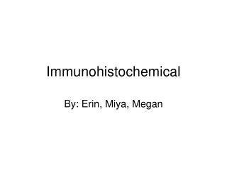
Immunohistochemical By: Erin, Miya, Megan
Definitions • Immunoflurescence- lab technique to identify antigens or antibodies • Immunohistochemical staining- detection of antigens in tissues • Immunocytochemistry- detection of antigens in cultured cells
How is it Performed • Immunofluorescence is a laboratory technique. The exact technique may vary depending on the specific antibody that is being investigated and between different laboratories. • In general, cells, tissue, or some other substance is placed onto microscope slides. A small amount of sample containing antibodies (typically serum, a liquid portion of blood) is placed over the cells or tissue, allowing the antibodies that are specific for the particular tissue or cellular antigens to bind. • The serum is washed away, and a second antibody that binds to human antibodies (often made in another animal species such as rabbits or goats) is applied to the slide. This second antibody has a fluorescent dye chemically linked to it. If the person's [or animal’s] serum has antibodies that bind to the tissue or cells, a bright fluorescence can be seen by use of a special microscope. • http://www.nlm.nih.gov/medlineplus/ency/article/003521.htm
Why do this method? • We use this to detect the antigen/antibody reaction within tissues and cells. • It determines specific distribution and localization of this complex.
A Visual Explanation Antibody Staining There are many methods that use the exquisite sensitivity of antibodies to locate a cellular component or a specific molecule (a tissue antigen). Here we focus on the two-step method where the primary antibody is generated against the molecule of interest. The secondary antibody is raised to recognize the primary antibody, and has a fluorescent tag as shown in the drawing on the right http://www.wellesley.edu/Biology/Concepts/Html/antibodystaining.html http://molpharm.aspetjournals.org/cgi/content/full/54/1/78/F2
Interpretation Immunohistochemistry results from mouse muscle tissue. The dystrophin antibody has bound to the dystrophin protein which is in abundance at the sarcolemma of each muscle fibre. The structure in the top right corner with a high amount of dystrophin is likely a nerve. http://resources.yesican-science.ca/polar_science2006/week7_lab.pdf
Interpretation • “In immunohistochemical techniques, there are several steps prior to the final staining of the tissue antigen, and many potential problems affect the outcome of the procedure… Patience is required to solve these problems.” http://www.piercenet.com/Proteomics/browse.cfm?fldID=F95B91A9-3DC1-4B56-8E8D-59CA044A8BA7
References • http://www.nlm.nih.gov/medlineplus/ency/article/003521.htm • http://www.wellesley.edu/Biology/Concepts/Html/antibodystaining.html • http://molpharm.aspetjournals.org/cgi/content/full/54/1/78/F2 • http://resources.yesican-science.ca/polar_science2006/week7_lab.pdf • http://www.piercenet.com/Proteomics/browse.cfm?fldID=F95B91A9-3DC1-4B56-8E8D-59CA044A8BA7