Download
1 / 32

E N D
1. ALGORITHMIC IMMUNOHISTOCHEMICAL DIAGNOSIS OF UNDIFFERENTIATED TUMORS & METASTATIC CARCINOMAS Mark R. Wick, M.D.
2. CURRENT DILEMMAS INDIAGNOSTIC IMMUNOHISTOLOGY Ever-increasing number of commercial reagents for use in evaluation of human specimens; as of 12/02, there were over 2000 such antibodies listed in the Linscott catalogue
Many confusing entries in the pathology literature, vis-a-vis the �specificity� (and implied usefulness) of a great many antibody reagents
3. �SPECIFICITY� OFIMMUNOHISTOLOGIC REAGENTS Related, but not identical, to classical definition given by Galen & Gambino:
Spec = True negatives/True negatives + False positives
In reality, �specificity� of immunohisto-logical reagents must be evaluated in many restricted and well-defined contexts
Hence, �specificity� is a relative term in this applied clinical setting
4. THE INTEGRATED APPROACH TO IMMUNOHISTOLOGY: USING �NON-SPECIFIC� REAGENTS TO ADVANTAGE One is faced with an �either-or� decision in diagnostic immuno-histology; either use no reagents at all (since none is �absolutely� specific in the most stringent terms), or use them in particular ways that are dictated by relative antibody specificities
The latter approaches do not �waste� information that is potentially gleaned from �non-specific� antibodies, and at the same time safeguard the user against erroneous interpretations
5. INTEGRATED ALGORITHMIC IMMUNOHISTOLOGY OF MALIGNANT TUMORS:NECESSARY CONDITIONS The user must control and standardize the processing of all tissues in his or her laboratory (e.g., fixative; fixation times; microwave-mediated antigen �retrieval�)
The user must personally define optimal antibody concentrations rather than accept the recommendations of manufacturers
Data must be accrued on spectra of reactivity for all antibodies used in the laboratory, either �in-house� or by adopting the EXACT methods of someone else who has already done this type of reagent analysis
6. INTEGRATED ALGORITHMIC IMMUNOHISTOLOGY OF MALIGNANT TUMORS:NECESSARY CONDITIONS (CONT.) Data on antibody performance must be used as the substrate for formal statistical analysis of specificity & sensitivity in well-defined diagnostic settings, producing a �relative values� system
The sequence of interpretation of panels of immunostains is governed by their relative statistical values within each predefined setting, moving from highest to lowest predictive values
Discrete morphological categories must be established within which the statistical data are applied; e.g., small round-cell tumors, large polygonal-cell tumors, etc.
7. POTENTIALLY MORPHOLOGICALLY-INDETERMINATE TUMORS: GENERIC CATEGORIES Small round-cell tumors
Large polygonal-cell tumors
Spindle-cell tumors
Pleomorphic tumors
Hematopoietic tumors (in subsets of the first two categories)
8. ALGORITHMIC IMMUNOHISTOLOGY: CHOICE OF REAGENTS Panels of antibodies in each algorithm are dependent upon the generic classification of the tumor under study
Several antibodies appear in more than one algorithm, but their places in the relative sequences of interpretation (or the diagnoses that positivity yields) differs from one setting to another
New antibodies may be substituted for old ones or used to supplement existing reagents in all algorithms
9. KEY POINTS IN ALGORITHMIC IMMUNOHISTOLOGY Consistency of results is totally dependent upon consistency of METHODOLOGY
In order to adopt the algorithms of another investigator, his or her methods MUST be followed down to the last detail or differences in antibody performance will result
10. POTENTIALLY UNDIFFERENTIATED SMALL CELL TUMORS Small cell squamous carcinoma
Small cell neuroendocrine carcinoma
Malignant melanoma
Lymphoma
Granulocytic sarcoma
Neuroblastoma
Primitive neuroectodermal tumor
Rhabdomyosarcoma
12. POTENTIALLY UNDIFFERENTIATED LARGE-CELL TUMORS Poorly-differentiated squamous cell carcinoma
Adenocarcinoma
Malignant melanoma
Malignant lymphoma
Granulocytic sarcoma
Epithelioid-cell sarcomas
Epithelioid sarcoma
Clear cell sarcoma
Alveolar soft part sarcoma
Epithelioid malignant peripheral nerve sheath tumor
Epithelioid leiomyosarcoma
14. POTENTIALLY UNDIFFERENTIATED SPINDLE-CELL TUMORS Sarcomatoid squamous cell carcinoma
Spindle-cell malignant melanoma
Leiomyosarcoma
Fibrosarcoma
Malignant fibrous histiocytoma
Malignant peripheral nerve sheath tumor
Angiosarcoma
16. POTENTIALLY UNDIFFERENTIATED PLEOMORPHIC TUMORS Sarcomatoid squamous cell carcinoma
Pleomorphic malignant melanoma
�Dedifferentiated� sarcomas
Malignant fibrous histiocytoma
Malignant peripheral nerve sheath tumor
Angiosarcoma
18. ANTIBODIES USED IN EVALUATION OF METASTATIC CARCINOMAS OF UNKNOWN ORIGIN Keratin & Epithelial membrane antigen
Prostate-specific antigen
Thyroglobulin
Thyroid transcription factor-1
Gross cystic disease fluid protein-15
S100 protein
Carcinoembryonic antigen
CA19.9
CA-125
Placental alkaline phosphatase
CD15
Estrogen & Progesterone receptor proteins
19. ANTIBODIES APPLIED TO THE STUDY OF SMALL ROUND-CELL TUMORS KERATIN S100
EMA HMB45
CD45 MART-1
CD15 FLI-1
VIMENTIN SYN
DES CD56
MSA CD99
MYOGENIN
20. ANTIBODIES APPLIED TO THE STUDY OF LARGE POLYGONAL-CELL TUMORS KERATIN S100
VIMENTIN HMB45
BER-EP4 MART-1
CD45 CEA
CD15 THROMBOMODULIN
MOC31 CALRETININ
CD117
21. REAGENTS APPLIED TO THE STUDY OF SPINDLE-CELL TUMORS KERATIN S100 PROTEIN
EMA CD56
VIMENTIN CD31
DESMIN CD34
MSA CD57
THROMBOMODULIN CD99
22. REAGENTS APPLIED TO THE STUDY OF PLEOMORPHIC TUMORS KERATIN HMB45
EMA MART-1
VIMENTIN THROMBOMODULIN
DESMIN CD31
MSA CD34
S100 PROTEIN CD99
23. REAGENTS USED IN PARAFFIN SECTION PHENOTYPING OF HEMATOPOIETIC TUMORS CD15 CD79A
CD20 LYSOZYME
CD30 MYELOPEROXIDASE
CD43 CD3
CD45 CD5
CD68 CD163
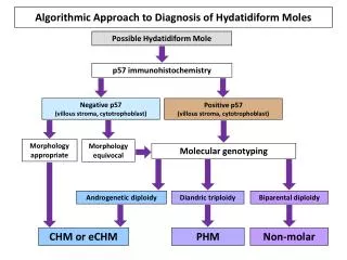
24. DETERMINATION OF ORIGIN FOR CARCINOMAS OF OCCULT OR CLOSELY-APPOSED SITES Use of immunohistochemistry in this setting relies on use of antigen �catalogues� for a variety of human carcinoma types
Antigens are grouped into �inclusionary� & �exclusionary� categories with respect to each tumor type, and arranged algorithmically in order of relative predictive values
Resulting antigen profiles constitute protein �fingerprints� of individual carcinoma types, which allow for their discrimination from one another
Few tumor-specific markers are available; hence, broad antibody panels are used
25. EXAMPLES OF CHARACTERISTIC IMMUNOPHENOTYPES OF SELECTED CARCINOMAS Breast: CK+; GCDFP15+; S100+/-; CEA-; ER+/-; CA125-; CA19.9-; PLAP-; EMA+; TTF-1-
Lung: CK+; GCDFP15-; S100-; CEA+; ER-; CA125-; CA19.9+/-; PLAP+/-; EMA+; TTF-1+
Kidney: CK+; GCDFP15-; S100-; CEA-; ER-; CA125-; CA19.9-; PLAP+/-; EMA+; TTF-1-
Adrenal: CK-; GCDFP15-; S100-; CEA-; ER-; CA125-; CA19.9-; PLAP-; EMA-; TTF-1-
Ovary: CK+; GCDFP15-; S100+/-; CEA-; ER+/-; CA125+; CA19.9+/-; PLAP+/-; EMA+; TTF-1-
Germ Cell: CK+/-; GCDFP15-; S100-; CEA-; ER-; CA125-; CA19.9-; PLAP+; EMA-; TTF-1-