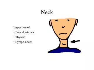
neck
neck. Lu tingting Regional anatomy department. brief. Muscle Vessel. Review. Fascia and fasical space. division of the neck. Thyroid and parathyroid glands. Root of the neck. Lymphatics of the neck. review. anterior. posterior. platysma. platysma. Sternoclei- domastoid.


neck
E N D
Presentation Transcript
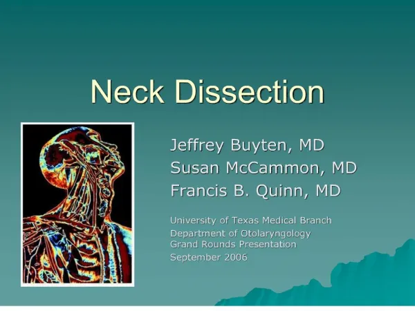
neck Lu tingting Regional anatomy department
brief Muscle Vessel Review Fascia and fasical space divisionoftheneck Thyroid and parathyroid glands Root of the neck Lymphatics of the neck
review anterior posterior
platysma Sternoclei- domastoid
Infrahyoid muscles thyrohyoid Omohyoid (superior and inferior belly) sternothyroid sternohyoid
stylohyoid digastric (anterior and posterior belly) Suprahyoid muscles mylohyoid
Thyroid gland Parathyroid glands
Hyoid bone(between c3 and c4) Thyroid cartiliage Cricoid cartilage(c6) Crico-thyroid membrane Tracheal cartilage (“c”)
mastoid Hyoid bone(between c3 and c4) Thyroid cartiliage Laryngeal prominence Cricoid cartilage(c6) scapula sternum
scalenus Vertebral column
Artery : Common carotid artery( upper border of thyroid cartilige level) internal and external carotid artery Vein: Internal and external jugular vein
artery Internal jugular vein External jugular vein
artery Common carotid artery Subclavian artery (Anterior and middle scalenus) Brachiocephalic artery
Internal carotid artery External carotid artery Carotid sinus
vein Internal jugular vein External jugular vein Anterior jugular vein
Analysis Give drug Parenteral nutrition (give nutrition from the recirculating system not digestive system ) Subclavian vein
Superificial fascia: Skin ,platysma Anterior jugular vein ,External jugular vein Nerve:cervical branch of facial nerve(platysma) cervical nerve Cervical fascia※ ( Investing layer Pretracheal layer Prevertebral layer)
pretracheal fascia Infrahyoid m. trachea esophagus thyroid Internal jungular vein Pretracheal layer s.c.m Commoncarotid a. Carotid sheath Vagus n. Buccopharyngeal fascia scalenus Investing layer Prevertebral layer Trapezius
carotid sheath Posterointernal-common and internal carotid artery Anteroexternal-internal jugular vein Between and behind them-vagus nerve anterior posterior
recurrent laryngeal nerve sympathetic trunk Vagus nerve
Fascial space: Pretracheal space Retropharyngeal space Fascial space within Pretracheal laryer
Buccopharyngeal fascia Prevertebe-al laryer Pretracheal space Retropharyn- geal space Fascial space within Prevertebral laryer
Pretracheal space: Thyroid ima artery, Inferior thyroid vein and thymus gland in baby Retropharyngeal space: Fascial space within Pretracheal laryer: cervical tuberculosis become axillary abscess and transfer to mediastinum
trapezius Nape Neck ※
s.c.m Anterior triangle Posterior triangle
Anterior belly of digastric m. Hyoid bone Superior belly of omohyoid m.
Submandibular triangle-submandibular gland Submental triangle-submental gland Carotid triangle-carotid sheath
division of the neck Anterior triangle Suprahyoid region: submental triangle submandibular triangle Infrehyoid region: muscular triangle※ carotid triangle※ Posterior triangle
muscular triangle Position Neighborhood Artery Vein Nerve Thyroid gland Superior laryngeal nerve Recurrent laryngeal nerve
SHAPE: H-shaped, isthmus, lateral lobes and pyramidal lobe 50%
POSITION: • Two lateral lobes:at lower part of larynx and anterolateral sides of upper part of trachea • Isthmus: anterior to 2nd--4th tracheal cartilages • Upper pole:mid-point of thyroid cartilage • Lower pole:6th tracheal cartilage
COVERING OF THE THYROID GLAND THYROID SHEATH (false capsule): formed by pretracheal fascia FIBROUS CAPSULE (true capsule): external membrane of thyroid gland
CAPSULE-SHEATH SPACE : between false and true capsules, contains loose connective tissue, blood vessels, nerves and parathyroid gland. SUSPENSORY LIG. OF THYROID GLAND: formed by false capsule, fix the gland at larynx and trachea, and moves up and down when swallowing
Neighborhood: Anterior:superficial fascia(skin,platysma…) investing fascia (s.c.m. infrahyoid muscles ) pretracheal fascia
posteromedial:trachea, esophagus, recurrent laryngeal nerve posterolateral: carotid sheath(common carotid artery, internal jugular vein and vagus nerve) cervical sympathetic trunk behind pretracheal fascia
CLINICAL SIGNIFICANCE tracheal compression-dyspnea(can not breath) esophageal compression-dysphagia(can not swallow) compression of recurrent laryngeal nerve-hoarse voice sympathetic trunk-Horner syndrome
ARTERIES OF THYROID GLAND • Superior thyroid artery: arise from the anterior aspect of the origin of the external carotid a. • Inferior thyroid artery :branch of thyrocervical trunk • Thyroid ima artery (10%):arise from brachiocephalic trunk of aortic arch
VEINS OF THYROID GLAND Superior thyroid vein:accompanied by homonymous artery and joins internal jugular vein. Middle thyroid vein: one, or two, three, or absent; joins internal jugular vein which is thin, preventing bleeding or air embolus when ligation Inferior thyroid vein:joins brachiocephalic veins in front of trachea. Plexus thyroideus impar:formed by anastomosis of branches of two inferior thyroid veins with tributaries of isthmus in front of trachea.
NERVES RELATED TO THYROID GLAND Superior laryngeal nerve Internal branch:distribute in the laryngeal mucous membrane above glottis vocalis , accompanying the superior laryngeal artery External branch:goes inferioanteriorly accompanying the superior thyroid artery, separates with the artery at 1 cm distant to upper pole of lateral lobe to supply cricothyroid muscles and inferior pharyngeal constrictor